
SHARE Knowledge Hub
What are corneal irritants and how should they be controlled?
A chemical splash or airborne irritant can injure the eye. Identify the specific substance and route, prevent contact at source, keep immediate rinsing available, and obtain urgent medical assessment for a chemical in the eye.
Understanding What are corneal irritants and how should they be controlled?
A chemical splash or airborne irritant can injure the eye. Identify the specific substance and route, prevent contact at source, keep immediate rinsing available, and obtain urgent medical assessment for a chemical in the eye.
Why this matters at work
Identify the exact chemical, product, concentration, form and task rather than applying the same advice to every eye irritant. Distinguish vapour or dust exposure, liquid splash, pressure injection, hot material and high-speed impact. Consider normal use, filling, sampling, cleaning, maintenance and foreseeable failures. Use the current product information and competent assessment for the actual conditions. The example name benzoquinone does not establish a universal exposure limit or the severity of a particular injury.
Reduce the hazard at source through closed handling, shielding, pressure control and suitable methods. Teach correct use, cleaning, storage and defect reporting, and replace damaged protection under the real programme. Check use in actual work, including cleaning and sampling. Do not approve a named model or infer protection against a chemical from appearance alone. Record the selection rationale and review after changes or exposure events. PPE does not establish that the remaining process or emergency arrangements are adequate.
How organisations manage this in practice
Prevent exposure through elimination or substitution where practicable, then suitable closed handling, source control, shielding, controlled pressure and safe methods. Select compatible eye and face protection for the residual mechanism, without relying on PPE as the only barrier. Plan accessible immediate irrigation and urgent medical help before work begins. Record the task, controls, checks and limitations. GB and NI COSHH use separate instruments. Diagnosis and individual treatment remain clinical decisions.
Review why the release occurred: blockage, trapped pressure, fittings, disconnection method, shielding and equipment condition. Select source and engineering controls before reviewing residual eye or face protection for the mechanism. Check whether other people or nearby surfaces were contaminated and manage access and cleanup competently. Record factual observations, task conditions and corrective evidence, keeping clinical information confidential. Reuse and return to work need separate accountable decisions. This example provides neither a pressure-release procedure nor a finding about a specific protective product.
Arrange suitable immediate and sustained irrigation capability and trained assistance through the competent local assessment. Check equipment instructions and a risk-based maintenance programme, including relevant water hygiene and winter conditions. Keep the affected task restricted if essential emergency precautions are missing. Record the gap, owner, temporary restrictions and verification. Test access and response through safe exercises without chemical exposure. The example concerns readiness and does not replace product-specific professional first-aid advice or determine compliance of a particular installation.
Keep the emergency eye route beside the substance-specific task
For a suspected corrosive eye exposure, use Chemical Eye Injuries and the first-response section immediately. Keep rinsing while urgent help is arranged; the eye-washing minimum is not a signal that suspected-burn treatment is complete. A high-speed particle, a chemical splash and a mixed powder exposure need their respective urgent assessment routes, not one assumption of harmless dust.
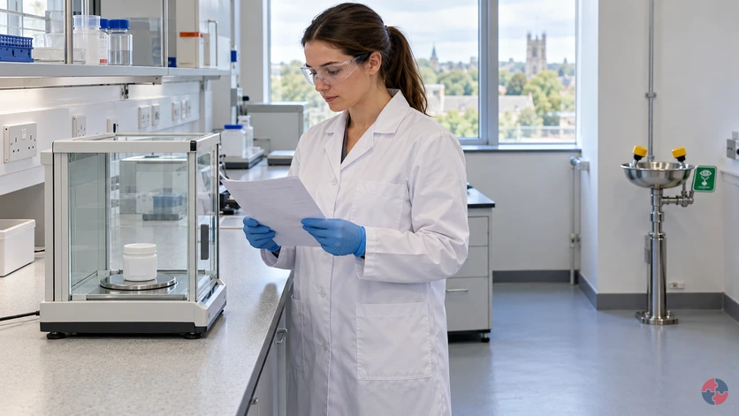
A balance draught shield is not verified containment
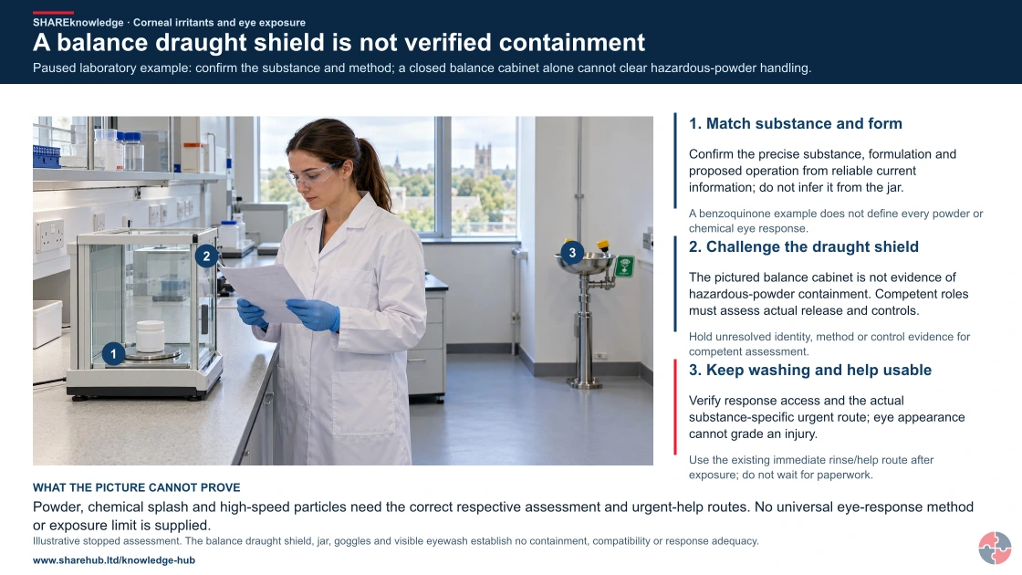
Full written explanation
Paused laboratory example: confirm the substance and method; a closed balance cabinet alone cannot clear hazardous-powder handling.
- Match substance and form. Confirm the precise substance, formulation and proposed operation from reliable current information; do not infer it from the jar.
- Challenge the draught shield. The pictured balance cabinet is not evidence of hazardous-powder containment. Competent roles must assess actual release and controls.
- Keep washing and help usable. Verify response access and the actual substance-specific urgent route; eye appearance cannot grade an injury.
Change the pathway
- Keep the task specific. A benzoquinone example does not define every powder or chemical eye response.
- Do not start open handling. Hold unresolved identity, method or control evidence for competent assessment.
- Urgent care has priority. Use the existing immediate rinse/help route after exposure; do not wait for paperwork.
Evidence check: Powder, chemical splash and high-speed particles need the correct respective assessment and urgent-help routes. No universal eye-response method or exposure limit is supplied.
Full clinically sourced urgent eye-response guidance and substance-specific context
Primary references: HSE: COSHH source controls (GB) · NHS: eye injuries and urgent care · NHS: acid and chemical burns
Open the complete HD image (new tab)Download image
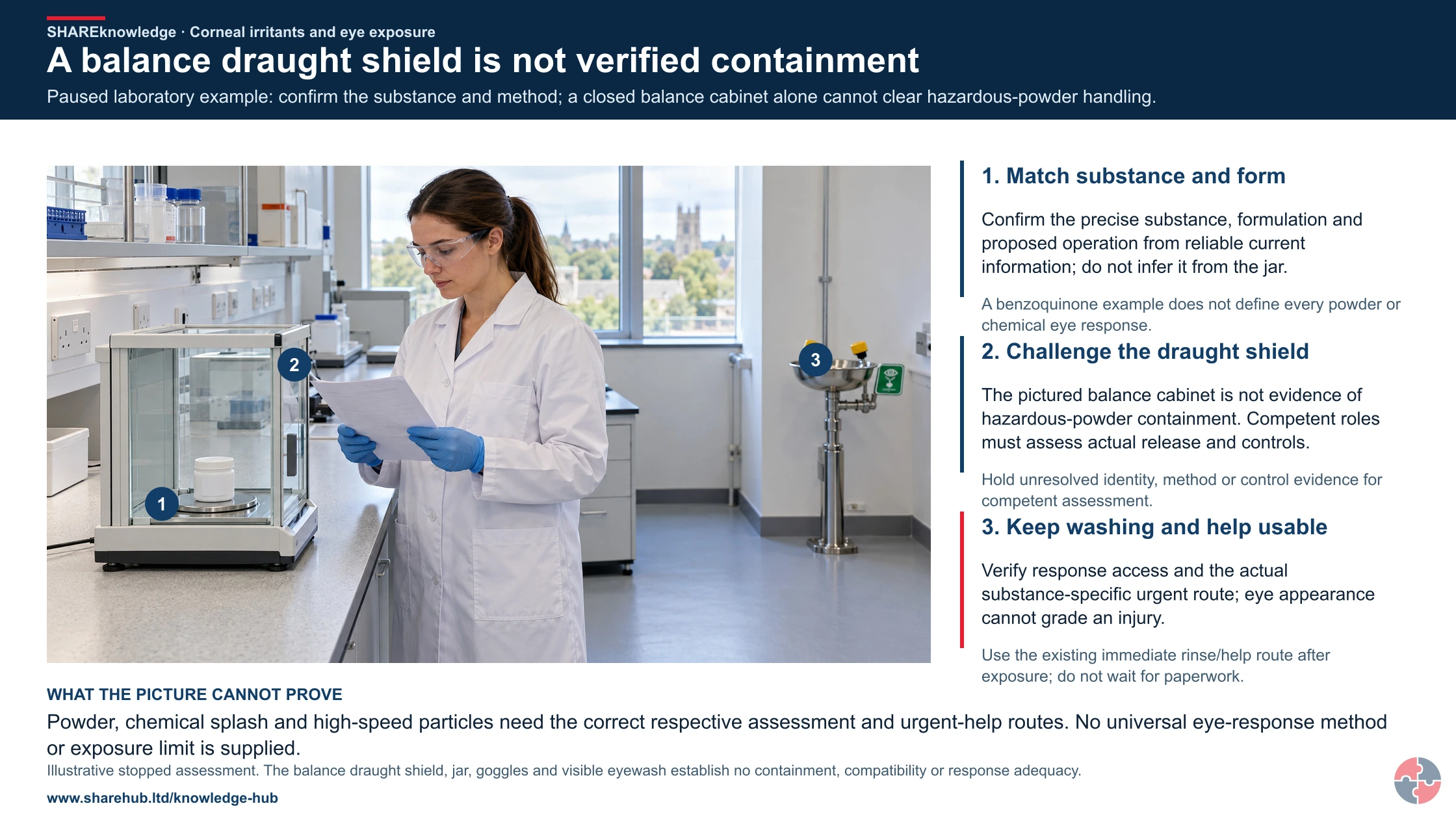
Full clinically sourced urgent eye-response guidance and substance-specific context
Primary references: HSE: COSHH source controls (GB) · NHS: eye injuries and urgent care · NHS: acid and chemical burns
For a distinct benzoquinone example, consider a small laboratory weighing task. Confirm the precise substance and formulation from reliable current information, avoid open transfer where practicable, assess airborne powder and hand-to-eye contact, and have the competent laboratory owner verify containment and protection. Do not assume a general leisure-cleaning example establishes the controls, or treat foreign technical guidance as UK law.
Laboratory example: benzoquinone powder transfer
Illustrative example. A laboratory proposes weighing a benzoquinone-containing material using an open bench method.
Decision. The laboratory assessment owner verifies identity and task hazards, considers a contained method and checks control and response capability before release.
Evidence to check. Actual formulation, task and exposure routes, suitable containment and eye protection, trained users and accessible water.
Hold or escalation point. Hold open handling if identity or suitable task controls are unresolved; use the urgent eye route after exposure.
Follow-up. Check the revised method in use and keep the laboratory assessment distinct from incidental dust or cleaning guidance.
Keep the splash source and response in view
The corneal surface inset explains location only. Prevention, accessible response facilities and substance-specific medical advice remain the practical priorities.
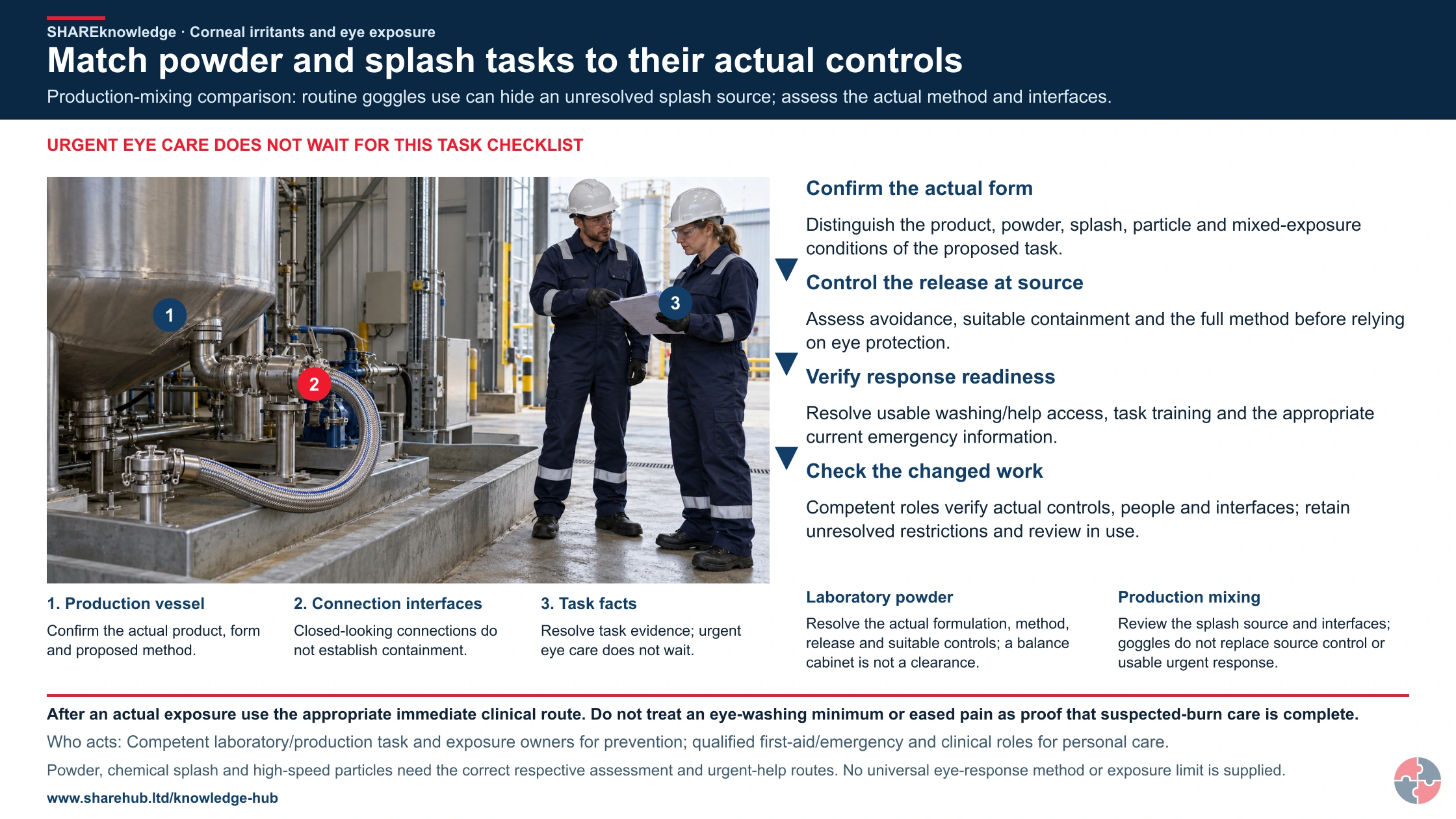
Match powder and splash tasks to their actual controls
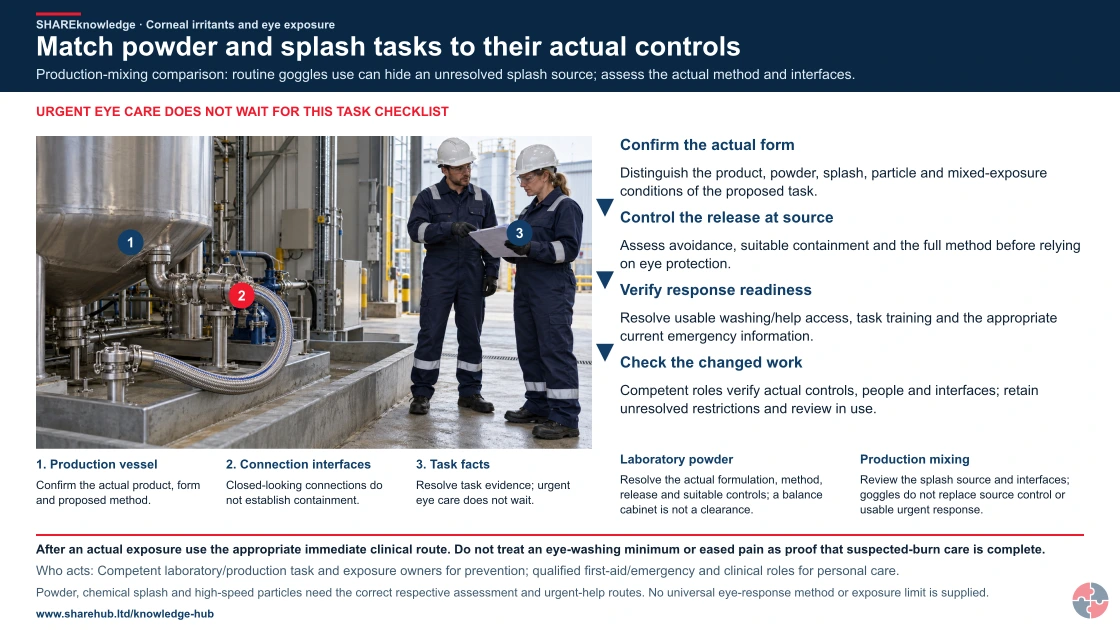
Full written explanation
An illustrative stationary production-equipment review shows a vessel, closed hose connections and two uninjured colleagues discussing inert task facts. Picture key: 1 identifies the production vessel; 2 identifies the connection interfaces; 3 identifies the task facts. The vessel prompts confirmation of the actual product, physical form and proposed method. The hose and couplings prompt questions about the actual release source, suitable containment and changed interfaces. Their appearance does not establish contents, chemical identity, flow, valve state, compatibility, containment performance or permission. This picture shows no mixing, powder release, splash, exposure or response demonstration. The complete prevention review keeps its original order: confirm the actual form, control release at source, verify response readiness and check changed work. Those independent headings are unnumbered; their arrows connect prevention checks only. Urgent personal care remains separate from task paperwork or investigation. The laboratory-powder and production-mixing contexts require their respective assessment and urgent-help routes; the picture supplies no universal eye response, numerical limit, eyewear selection or operating method. Read the complete source for the existing clinical, local-care and national qualifications.
Production-mixing comparison: routine goggles use can hide an unresolved splash source; assess the actual method and interfaces.
- Confirm the actual form. Distinguish the product, powder, splash, particle and mixed-exposure conditions of the proposed task.
- Control the release at source. Assess avoidance, suitable containment and the full method before relying on eye protection.
- Verify response readiness. Resolve usable washing/help access, task training and the appropriate current emergency information.
- Check the changed work. Competent roles verify actual controls, people and interfaces; retain unresolved restrictions and review in use.
When the evidence does not fit: After an actual exposure use the appropriate immediate clinical route. Do not treat an eye-washing minimum or eased pain as proof that suspected-burn care is complete.
Who acts: Competent laboratory/production task and exposure owners for prevention; qualified first-aid/emergency and clinical roles for personal care.
Changed workplace
Laboratory powder: Resolve the actual formulation, method, release and suitable controls; a balance cabinet is not a clearance.
Production mixing: Review the splash source and interfaces; goggles do not replace source control or usable urgent response.
Evidence check: Powder, chemical splash and high-speed particles need the correct respective assessment and urgent-help routes. No universal eye-response method or exposure limit is supplied.
Full clinically sourced urgent eye-response guidance and substance-specific context
Primary references: HSE: COSHH source controls (GB) · NHS: eye injuries and urgent care · NHS: acid and chemical burns
Open the complete HD image (new tab)Download image
Full clinically sourced urgent eye-response guidance and substance-specific context
Primary references: HSE: COSHH source controls (GB) · NHS: eye injuries and urgent care · NHS: acid and chemical burns

Identify the substance
Confirm the actual product and assessed response information; do not generalise from one example irritant.
Prevent contact
Assess containment, splash control and compatible eye/face protection for the task.
Response and help
Keep appropriate washing and assistance accessible and follow the clinically sourced response on this page.
No anatomical picture can grade the injury or justify delaying appropriate medical help.
What are the key facts about corneal irritants?
The key facts are that eye exposure can cause immediate pain, rapid chemical injury, mechanical abrasion or delayed inflammation, so prevention and fast response both matter.
Chemical eye exposure can deteriorate quickly. Significant splashes, corrosive or irritant exposure, continuing pain, blurred vision, light sensitivity, embedded material or difficulty opening the eye should be managed through workplace first-aid arrangements and medical assessment. The exposure task should be stopped and reviewed before similar work continues.
The cornea is highly sensitive
Small injuries can be very painful and may affect vision.
Liquids are not the only problem
Dusts, powders, aerosols, fumes, vapours and flying particles can also injure the eye.
Corrosives are higher consequence
Acids and alkalis can cause serious injury quickly, especially if irrigation is delayed.
PPE must match the hazard
Safety glasses may not stop splashes, powders or high-pressure liquids reaching the eye.
Eyewash has to be usable
It must be accessible, clean, understood and available when the task creates foreseeable eye exposure.
Near misses matter
A splash on goggles, poor decanting method or blocked eyewash point is evidence that controls need review.
What are corneal irritants?
Corneal irritants are substances or particles that can inflame or damage the cornea and surrounding eye tissues.
The cornea is the transparent front layer of the eye. The CUH corneal-abrasion guidance explains that the cornea contains many nerves and a scratch can be very painful; grit can scratch it.
Workplace eye exposure can be chemical, mechanical or mixed. A solvent splash may irritate the eye; a corrosive alkali such as ammonium hydroxide can penetrate rapidly and cause permanent injury, as described by UKHSA’s ammonia overview. A dusty powder may scratch the surface and chemically irritate it; a fume or vapour may cause watering and inflammation.
Benzoquinone is included as an example because it is a reactive irritant chemical. The practical lesson is not that every workplace has benzoquinone; it is that the exact substance, concentration, physical form and task determine the eye-hazard controls.
| Exposure type | Examples | Why it matters |
|---|---|---|
| Irritant liquid | Cleaning chemicals, solvents, oils and metalworking fluids. | Can cause redness, pain and inflammation. |
| Corrosive chemical | Strong acids, alkalis, caustic cleaners and cement slurry. | Can cause rapid tissue damage. |
| Powder or dust | Cement, lime, powders, grit, wood dust and process dust. | May scratch and chemically irritate the eye. |
| Mist or aerosol | Sprays, pressure washing, disinfectants and coating work. | Can bypass ordinary glasses and reach the eye. |
| Fume or vapour | Reactive chemicals, welding by-products and volatile irritants. | Can cause watering and irritation. |
| Flying particle | Grinding, cutting, drilling and compressed-air misuse. | Can cause abrasion or embedded foreign body. |
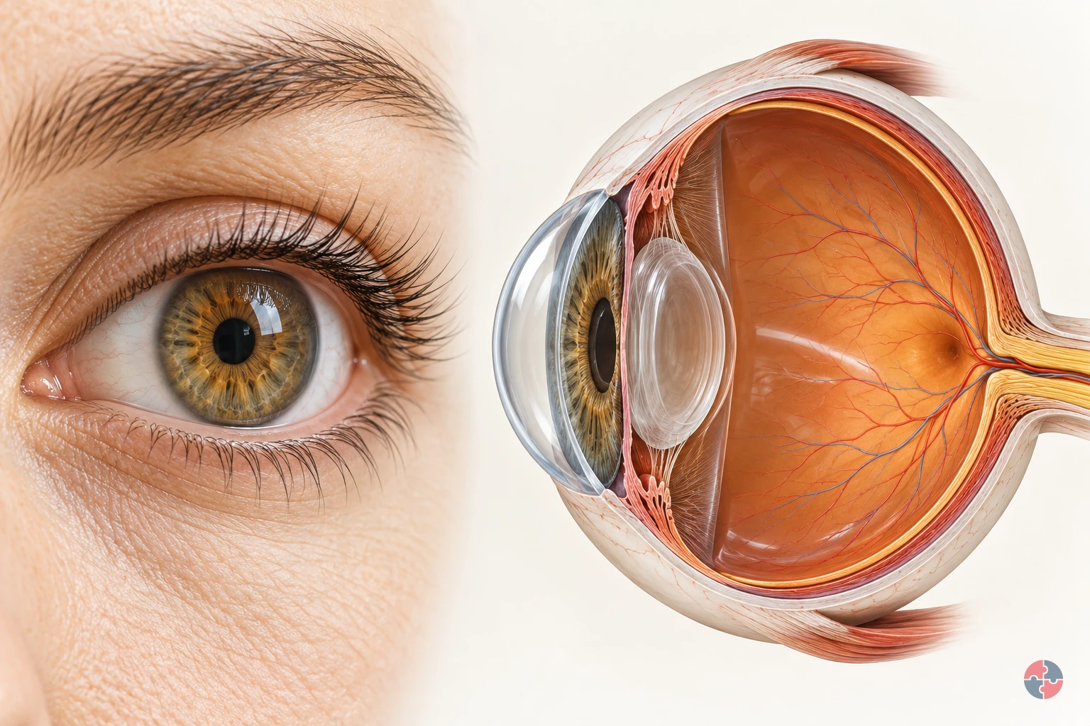

How does the eye turn light into vision, and how do workplace hazards injure it?
Interactive anatomy · Eye function and injury · Updated 2026-10-23
Read all anatomy and process explanations without the viewer
Educational illustration, not a diagnostic symptom checker or a reproduction of an individual’s vision. No acuity/brightness challenge, flashing screen, camera permission, eye-drop prescription, patching instruction, lens-removal manoeuvre or home-neutralisation exercise. Ordinary DSE comfort is separate from trauma; eyesight correction is separate from hazard protection.
Eyelids
Blinking spreads tears over the front of the eye. The eyelids pass over the exposed front of the eye when a person blinks. Their movement spreads the tear film, helping keep that surface moist and smooth. In the surface view, use the lid margins to locate the area they cover. This surface-maintenance role supports the optical pathway; the lid does not carry visual signals to the brain. Moorfields Eye Hospital NHS Foundation Trust: Dry eye: the tear film
Tear film
A thin tear layer keeps the surface moist and smooth. A thin film of tears covers the front of the eye. It lubricates the surface and contributes to a smooth boundary through which light passes. Blinking redistributes this film. Its thickness is enlarged in the teaching view so its position can be seen; it is a surface layer rather than a pool of liquid inside the eyeball. Moorfields Eye Hospital NHS Foundation Trust: Dry eye: the tear film
Cornea
The clear front surface bends incoming light. The transparent cornea forms the curved front of the eye, providing an outer protective boundary and bending incoming light. It works with the lens to focus the light towards the retina. Follow the rays through the front curve in the cutaway, then compare its location with the tear film lying over the surface. Moorfields Eye Hospital NHS Foundation Trust: What makes up your eye
Iris
The coloured ring controls the pupil opening. The iris is the coloured ring around the pupil. Its muscles change the size of the opening and so control how much light can enter the eye. Locate the ring separately from the transparent lens behind it. This light-entry role is different from the focusing work performed by the cornea and lens. Moorfields Eye Hospital NHS Foundation Trust: What makes up your eye
Pupil
An opening lets light enter the eye. The pupil is the central opening in the iris through which light passes. It looks dark when viewed from the front because it opens into the inside of the eye. It is not a solid black disc. In the cutaway, follow the route through this opening towards the transparent lens behind it. Moorfields Eye Hospital NHS Foundation Trust: What makes up your eye
Lens
The clear lens helps focus light on the retina. The transparent lens sits behind the iris and adds to the focusing action of the cornea. In a living eye its shape can change to help focus on objects at different distances. Use the cutaway to locate the lens between the pupil opening and the retina; the simplified ray diagram is not an eyesight test. Moorfields Eye Hospital NHS Foundation Trust: What makes up your eye
Retina
The light-sensitive layer lines the back of the eye. The retina is the light-sensitive tissue lining the inside of the back of the eye. Light focused by the cornea and lens reaches this layer, where specialised cells begin converting it into electrical information. Its position against the inner wall connects the whole-eye view with the separate enlarged photoreceptor inset. Moorfields Eye Hospital NHS Foundation Trust: What makes up your eye · Oxford University Hospitals NHS Foundation Trust: Tackling vision loss: normal signal pathway
Photoreceptors
Specialised retinal cells begin the light-to-signal conversion. Photoreceptors are specialised cells in the retina that respond to light and begin the conversion into electrical signals. Locate the retinal lining first, then connect it with the enlarged cell inset. That inset explains a change of process and scale; it does not represent a separate organ or every cell involved in retinal processing. Oxford University Hospitals NHS Foundation Trust: Tackling vision loss: normal signal pathway
Optic nerve
Visual signals travel from the eye towards the brain. The optic nerve carries electrical information from the retina towards the brain. In the model, the symbol changes from a light ray to a neural signal at the retina to distinguish these processes. Locate where the nerve leaves the back of the eye. Light itself does not travel along the nerve as a beam. Oxford University Hospitals NHS Foundation Trust: Tackling vision loss: normal signal pathway
Brain destination
The brain processes the incoming visual signals. The brain processes the visual information arriving from the eyes, helping us make sense of what we see. This marker shows a destination outside the eyeball rather than a structure within it. Follow the optic-nerve route to the marker after locating the retina; the detailed pathways inside the brain are not depicted here. Oxford University Hospitals NHS Foundation Trust: Tackling vision loss: normal signal pathway
Open the complete process transcript
The process, step by step
1 · Refresh the surface
A blink carries a thin layer of tears across the exposed front of the eye. This tear film keeps the surface moist and helps create a smooth first surface for incoming light. Look at the eyelids and the enlarged tear layer together: the lid spreads the film rather than pushing tears into the eyeball. The surface view separates these structures so their relationship is easier to see. This is the preparation for the optical pathway that follows; a clear view depends on the condition of the front surface as well as the structures deeper inside. Moorfields Eye Hospital NHS Foundation Trust: Dry eye: the tear film
What to watch: Locate the eyelid margins and the thin tear layer in front of the cornea, using the surface view to separate them.
Why it matters: The refreshed film lubricates the front of the eye and helps keep its surface smooth. It supports the passage of light before the cornea and lens focus that light deeper inside.
Explore this step: Select the eyelids, then the tear film, and describe which structure moves and which surface it covers.
2 · Bend incoming light
The cornea is the transparent, curved front of the eye. Incoming light changes direction as it passes through this surface, beginning the focusing process. In the whole-eye cutaway, trace the illustrative rays from outside the eye towards the lens. Their bend shows the cornea doing optical work rather than acting only as a window. The cornea also forms part of the protective outer boundary, so its clarity and shape matter to seeing. The rays simplify the route through a healthy eye; they are not a measurement of an individual person’s focus or vision. Moorfields Eye Hospital NHS Foundation Trust: What makes up your eye
What to watch: Find the clear curved front of the eye and follow where the incoming light rays change direction as they pass through it.
Why it matters: Beginning to direct the light here allows the cornea and lens to work together. A transparent front surface lets light enter while forming part of the eye’s protective boundary.
Explore this step: Trace one ray from outside the cornea to the lens and identify the first place its direction changes.
3 · Focus towards the back
After passing through the cornea, light enters through the pupil, the opening in the coloured iris. The iris changes the size of that opening to control how much light enters. Behind it, the transparent lens works with the cornea to bring light towards the retina at the back. Follow the model from front to back and distinguish the pupil opening from the solid lens. In a living eye the lens can change shape to help focus at different distances. This simplified view explains the route and the relationship between the structures, rather than testing accommodation or eyesight. Moorfields Eye Hospital NHS Foundation Trust: What makes up your eye
What to watch: Identify the iris around the pupil opening, the transparent lens behind it and the light-sensitive retina lining the back of the eye.
Why it matters: Controlling light entry and focusing it are different jobs. Together they allow light to reach the sensitive tissue at the back in a useful pattern for the next stage of vision.
Explore this step: Name the opening light passes through, then the transparent structure it reaches next; follow the route to the retina.
4 · Convert at the retina
The retina is the light-sensitive tissue lining the back of the eye. Its photoreceptor cells respond to light and begin converting it into electrical information. Locate the retinal lining, then use the enlarged cell inset to connect the whole-eye view with this smaller-scale event. The inset makes the conversion easier to discuss without suggesting that the cells are that large inside the eye. At this point the lesson changes from the path of light to the path of a nerve signal. The model does not show every retinal cell or all the processing that takes place between them. Oxford University Hospitals NHS Foundation Trust: Tackling vision loss: normal signal pathway
What to watch: Locate the retina on the inner back wall, then connect it with the enlarged photoreceptor inset outside the whole-eye cutaway.
Why it matters: This conversion connects the eye’s optical system with its nervous system. The focused light pattern can now be represented by signals that travel from the retina towards the brain.
Explore this step: Point to where the light pathway ends and explain why the enlarged cells are an inset rather than another organ.
5 · Carry and interpret the signal
Visual information leaves the retina through the optic nerve and travels towards the brain. The brain processes this incoming information so that we can recognise and make sense of what we see. Follow the model’s neural-signal symbol from the back of the eye to the external brain marker, and compare it with the earlier light rays. The symbols represent different processes: a nerve carries signals, not a beam of light or a miniature picture travelling through a tube. The brain marker identifies the destination outside the eyeball; the detailed visual pathways within the brain are beyond this simplified model. Oxford University Hospitals NHS Foundation Trust: Tackling vision loss: normal signal pathway
What to watch: Find where the optic nerve leaves the back of the eye and trace its route towards the separate brain destination marker.
Why it matters: The earlier stages deliver and convert light; this stage carries the resulting information onwards. Vision therefore depends on the retina, its nerve connection and the brain working with the eye’s optical structures.
Explore this step: Compare the light-ray and neural-signal symbols, then explain what travels through the pupil and what travels through the optic nerve.
Read the practice situations and all answer explanations
Sources and how the model is made
The opening images are AI-generated anatomical illustrations. The interactive 3D models and labelled SVG diagrams use original procedural artwork. These teaching images are not medical scans or diagnostic tools. Source references support the learner explanations and do not grant artwork rights.
- Moorfields Eye Hospital NHS Foundation Trust: What makes up your eye — Normal cornea, iris, pupil, lens and posterior eye layout; use anatomy text selectively. No image copied.
- Moorfields Eye Hospital NHS Foundation Trust: Dry eye: the tear film — Blinking spreads the tear film, which lubricates the eye and supports a smooth surface. No treatment instructions copied.
- Guy’s and St Thomas’ NHS Foundation Trust: Corneal abrasion — A scratch/graze of the cornea and the distinction between a superficial injury and complications; no healing-time promise or eye treatment prescribed.
- NHS: Eye injuries — March 2026 guidance: immediate chemical irrigation and emergency help; high-speed/penetrating injury; severe pain or sight changes. Minimum rinsing time is not a clearance test.
- University Hospitals Dorset NHS Foundation Trust: Chemical injury to the eye — Possible surface/anterior-segment damage only. Printed review date November 2023 is overdue; current emergency actions rely on EY-S4, not this leaflet alone.
- HSE: Health risks from welding: arc-eye — Welding radiation can injure the cornea, cause delayed pain and harm other eye structures; no safe viewing time.
- HSE: Using the right type of PPE — Eye protection must match impact, splash, dust or radiation hazards and fit the task/user.
- Oxford University Hospitals NHS Foundation Trust: Tackling vision loss: normal signal pathway — Photoreceptors convert light to electrical signals, carried through the optic nerve to the brain. Research treatment content is not reused.
- NHS Organ Donation: About cornea donation — Injury or disease can leave the cornea cloudy/distorted and affect vision. Source is factual context, not donation advice in this lesson.
- University Hospital Southampton NHS Foundation Trust: Eye emergency department — Chemical injury, penetrating trauma and severe visual symptoms require emergency eye care. Local opening hours/contact instructions are not generalised.
How can a corneal irritant cause harm?
Severity depends on the actual chemical, concentration, route and time in contact; the example 1,4-benzoquinone can irritate the eye and inflame the cornea.
The NIOSH quinone entry lists eye irritation, conjunctivitis and keratitis for 1,4-benzoquinone. This is US toxicology context, not a UK workplace exposure limit or a diagnosis. Use the current product safety data sheet and HSE COSHH assessment to assess splash, mist, dust and transfer tasks. A worker with pain or visual change needs clinical assessment; do not label every red eye a benign irritant reaction.
Which forms and routes of exposure should be assessed?
The assessment should consider liquid splash, spray, dust, powder, vapour, fume, flying particles and contaminated hands or gloves.
Eye exposure can occur during routine work or during mistakes: decanting into the wrong container, opening a pressurised line, cleaning a blockage, pouring concentrate, using compressed air, removing contaminated gloves, leaning over a mixing vessel or touching the eye with contaminated fingers.
The task should be observed at the point of use. SDS information is important, but it rarely captures splash direction, worker posture, container height, ventilation, reaction heat, pressure, cleaning method or how people actually remove PPE.
Secondary contamination also matters. A worker can protect the eye during the task and then contaminate it afterwards by rubbing the face with dirty gloves or sleeves.
| Route or form | How it reaches the eye | Controls |
|---|---|---|
| Splash | Pouring, decanting, mixing, cleaning or pressure release. | Closed transfer, splash guards and goggles/face shield. |
| Spray or mist | Spraying, pressure washing or aerosol generation. | Enclosure, distance, ventilation and sealed eye protection. |
| Dust or powder | Scooping, tipping, cutting, sanding or dry sweeping. | Dust control, extraction and eye protection. |
| Vapour or fume | Volatile or reactive chemicals near the face. | Ventilation and suitable goggles where needed. |
| Flying particle | Grinding, drilling, cutting and impact work. | Machine guarding and impact-rated protection. |
| Hand-to-eye contact | Contaminated gloves or sleeves touch the face. | Hygiene, glove removal training and washing. |
What should happen if a chemical enters the eye?
Begin rinsing immediately with clean water and seek urgent medical help; do not delay irrigation for paperwork or a supervisor.
The NHS eye-injury guide advises plentiful clean water for at least 20 minutes, with emergency help for a strong chemical or serious symptoms. Keep the container or safety data sheet available for clinicians if safe to do so. Do not rub the eye, apply neutralising chemicals or treat the page as an ophthalmology protocol. Arrange an accessible eyewash point and practise the response before the task begins.
NHS eye-injury advice, reviewed 12 March 2026 says to continue rinsing while emergency help is arranged for a strong chemical, sight changes or other listed serious symptoms. Do not drive to A&E with an eye injury; arrange transport or an ambulance as needed. Follow the local emergency route outside England.
How should corneal irritants be controlled?
Corneal irritants should be controlled by preventing eye exposure before relying on eyewear alone.
The best controls reduce the chance of the substance or particle reaching the face at all. That may mean substituting a less irritating product, buying diluted product, using closed dispensing, reducing container height, fitting splash guards, improving extraction, changing nozzle pressure, using wet methods, eliminating compressed-air cleaning or segregating dusty work.
Eye protection remains important, but it must be selected for the exposure. Open safety glasses may be suitable for low-energy particles but weak for liquid splash. Chemical splash goggles may be needed for pouring concentrates. A face shield can protect the face but may need to be worn with goggles because liquid can travel behind it.
Controls should be checked in real work. If goggles fog, do not fit over prescription glasses, are scratched, are uncomfortable or are unavailable in the right size, workers may avoid them. The procurement and supervision system must solve those practical problems.
| Control area | Good practice | Common weakness |
|---|---|---|
| Substitution | Use less irritating or pre-diluted products where possible. | Same hazardous product used by habit. |
| Closed handling | Use pumps, dosing systems and sealed transfer. | Manual pouring creates splash risk. |
| Splash control | Use guards, trays, slow-pour methods and correct container height. | Workers lean over open containers. |
| Dust and particle control | Use extraction, wet methods and guards. | Dry sweeping or compressed air spreads particles. |
| Eye and face PPE | Match glasses, goggles and face shields to splash/impact risk. | Wrong type or poor fit. |
| Eyewash provision | Locate, maintain and train people in use. | Eyewash blocked, empty or too far away. |
How can the hierarchy of controls reduce eye exposure to corneal irritants?
Remove or substitute hazardous handling, enclose and contain splashes, use safe transfer procedures, and select eye/face PPE for residual risk.
For decanting a quinone solution, first ask whether a closed prefilled cartridge avoids the transfer. If not, assess a less hazardous compatible formulation, then use a closed pump and splash shield. Train workers and position eyewash within immediate reach. HSE eye-contact guidance and HSENI COSHH support prevention. Stop on a leaking connection, failed enclosure or inaccessible eyewash.
On a small screen, scroll this table sideways to read all columns.
How can the ERICPD hierarchy of controls reduce eye exposure to corneal irritants?

| Five-level / ERICPD | Control choice | Owner, check, failure and stop |
|---|---|---|
| Eliminate / eliminate | Use closed prefilled cartridge if feasible. | Buyer records decision; hold open decant if closed route works. |
| Substitute / reduce | Use compatible lower-hazard formulation after assessment. | Chemist approves; stop if new formulation hazards are unknown. |
| Engineering / isolate and control | Closed pump and splash shield. | Supervisor tests connections; stop on leak. |
| Administrative / discipline | Task COSHH, eyewash check and spill response. | Lead observes; stop if eyewash unavailable. |
| PPE / PPE | Suitable eye/face protection for residual splash. | Trainer checks fit; no work if protection fails. |
How can the ISO 45001 hierarchy of controls reduce eye exposure to corneal irritants?

| Five-level / ERICPD | Control choice | Owner, check, failure and stop |
|---|---|---|
| Eliminate / eliminate | Use closed prefilled cartridge if feasible. | Buyer records decision; hold open decant if closed route works. |
| Substitute / reduce | Use compatible lower-hazard formulation after assessment. | Chemist approves; stop if new formulation hazards are unknown. |
| Engineering / isolate and control | Closed pump and splash shield. | Supervisor tests connections; stop on leak. |
| Administrative / discipline | Task COSHH, eyewash check and spill response. | Lead observes; stop if eyewash unavailable. |
| PPE / PPE | Suitable eye/face protection for residual splash. | Trainer checks fit; no work if protection fails. |
HSE five-level hierarchy supplies the priority; ERICPD complements it. For quinone transfer, specify the pump, seals, connections, container support and shield for the actual chemical, concentration, temperature, pressure and splash path. A competent installer checks the assembled system under representative safe conditions and records acceptance before hazardous transfer. Keep manufacturer instructions, maintenance and defect records. Before each transfer and during use, check connection integrity, containment, shield position and accessible usable eyewash; recheck after container or setup changes. Leakage or failed protection stops transfer. Isolate safely, correct the cause and demonstrate the accepted arrangement before authorised restart. S100 containment and contamination guidance supports prevention; S102 eye/face protection guidance supports compatible protection selected for splash, dust, gas or vapour. Ordinary spectacles or a face visor alone may be insufficient; obtain competent selection advice for the actual exposure.
Which duties apply to corneal irritants?
GB COSHH or NI COSHH requires suitable task assessment and exposure control; emergency eye care remains clinician-led.
Use HSE COSHH for GB and HSENI COSHH for NI. The NHS advice supports immediate first response but is an England public-health page; follow local emergency care access in the work location. The NIOSH quinone sheet is hazard context, not UK law.
S100 is an October 2003 sheet; S102 also contains historical marking and standards references. Use their practical containment, hygiene and protection principles with current legal and equipment checks, rather than copying historic approval markings. NIOSH quinone entry was reviewed 30 October 2019; its US exposure limits are not UK limits.
How should corneal irritants appear in the risk register?
The risk register should describe the substance, eye-exposure route, task, people affected, controls and evidence.
A useful entry does not simply say eye injury. It should say whether the credible event is an alkali splash, dust abrasion, vapour irritation, spray mist, flying particle, contaminated glove contact or pressure-release incident. This makes controls specific enough to check.
| Scenario | Critical controls | Evidence |
|---|---|---|
| Pool chemical splash during dosing | Closed dosing, goggles/face shield, eyewash and training. | COSHH assessment and inspection. |
| Cement slurry in eyes | Wet cement control, splash goggles, gloves and eyewash. | Method statement and briefing. |
| Benzoquinone handling in laboratory | Small quantities, fume cupboard, goggles and spill plan. | SDS review and local rules. |
| Grinding particle injury | Guarding, impact eye protection and exclusion. | Tool inspection and PPE checks. |
| Aerosol disinfectant exposure | Spray control, ventilation and sealed eye protection where needed. | Task observation and controls. |
| Contaminated glove eye contact | Glove removal, hygiene and face-touch prevention. | Training and supervision. |
How could Riverside Leisure Ltd manage corneal irritant risks?

Riverside Leisure Ltd could manage corneal irritants by treating pool chemicals, cleaning products, aerosols and maintenance dusts as practical eye-exposure risks.
Riverside Leisure Ltd
Riverside Leisure Ltd is a leisure, events and holiday park business used by SHARE to show how health and safety controls work in customer-facing environments.
Riverside keeps a COSHH register for pool dosing chemicals, cleaning concentrates, descalers, disinfectants, aerosols, workshop products, grounds chemicals and maintenance dusts. Each assessment identifies the eye-exposure route: splash, mist, vapour, dust, hand-to-eye contact or flying particle.
Procurement favours closed dosing systems, pre-diluted products and containers designed to reduce splashing. Chemical stores keep eyewash, SDS access and segregation arrangements. Contractors are told which products and areas they may use, and higher-risk work such as pool plant maintenance is planned with permit-style controls.
Supervisors complete frontline checks on goggles, face shields, eyewash access, chemical labels, dilution systems, spill kits and welfare. Second-line HSSE checks review incident reports, near misses, training records, PPE replacement, first-aid stock and whether workers can explain what to do after a splash.
If an eye exposure or near miss occurs, Riverside records the substance, task, PPE, eyewash use, first-aid response and whether the worker received medical assessment. The learning is then used to amend the COSHH assessment, retrain the team, improve storage or change the product.
| Management element | How Riverside applies it | Evidence |
|---|---|---|
| Assessing | COSHH assessments identify eye route, substance form and task. | COSHH register and SDS records. |
| Analysing | Incidents and near misses are grouped by product and task. | Trend review and action tracker. |
| Implementing | Uses closed dosing, splash goggles, eyewash and clear storage rules. | Inspections and procurement records. |
| Training | Workers receive awareness, task and supervisor-level training. | Training matrix and briefings. |
| Checking | Supervisors and HSSE advisers verify PPE, eyewash and work methods. | Check sheets and audits. |
| Learning | Eye exposure events trigger product, method or PPE review. | Investigation records and updated assessments. |
What should each role learn and demonstrate?
Learning must match the decisions people make, from recognising danger to checking that controls work.
Briefings should tell people how to act, but attendance alone does not show competence. Assess the real decision or task, record who assessed it and revisit after a material change, failed control or incident. A local authorisation is task-specific; no universal external certificate is implied.
On a small screen, scroll this table sideways to read all columns.
| Role and learning | Demonstration and assessor | Record and reassessment trigger |
|---|---|---|
| Worker | Demonstrate transfer and immediate rinse; supervisor assesses. | Observation; repeat after spill. |
| Supervisor | Check enclosure, eyewash and SDS; hygienist observes. | Inspection; repeat on product change. |
| First aider | Practise irrigation and urgent referral; qualified trainer assesses. | Drill; repeat after failure. |
How does this topic support study and professional development?
Corneal irritants help learners apply COSHH, acute injury prevention, PPE selection and first-aid arrangements to a specific route of entry.
For NEBOSH General Certificate study, useful answers identify the substance, route into the body, likely harm, control hierarchy, PPE suitability, first-aid arrangements, training and review after incidents. Eye exposure scenarios are good tests of whether learners move beyond generic PPE answers.
For Diploma-level learning, the topic links toxicology, acute injury, occupational hygiene, substance classification, human factors, first-aid needs assessment and management-system assurance. For CPD, useful learning includes COSHH assessor training, chemical handling, PPE selection, first aid at work, incident investigation and supervisor inspections.
| Learning route | What to know | Practical application |
|---|---|---|
| NEBOSH General Certificate | COSHH, PPE, first aid and route of entry. | Explain controls for a chemical splash scenario. |
| NEBOSH Diploma | Substance classification, toxicology and assurance. | Evaluate whether controls are suitable and evidenced. |
| Supervisor CPD | How to check eyewash, PPE and work methods. | Run focused inspections. |
| First-aid CPD | Chemical eye exposure response and escalation. | Support timely irrigation and referral. |
| COSHH assessor CPD | Eye hazard assessment and control selection. | Improve task-specific assessments. |
How can a management system sustain effective decisions?
Give each hazard or support route an owner, a control, a check and a response to failure.
Connect product identity, transfer method, engineering control, eyewash readiness and urgent referral. An owner should sample actual work and corrective actions, not accept a policy or certificate as evidence that the controls work.
The free GB regulator guide HSE Managing for health and safety (HSG65) explains a plan–do–check–act approach: assign controls and owners, involve workers, check work as done, and correct failures. For Northern Ireland, use HSENI risk assessment guidance for the local duty. A certificate alone does not demonstrate that a control works.
What warning signs show corneal irritant risk is not controlled?
Warning signs include splashes on PPE, missing eyewash, unsuitable goggles, poor labelling, worker improvisation and repeated eye irritation reports.
Warning signs show how harm could occur. A worker pouring concentrate at face height, goggles fogging so they are removed, eyewash hidden behind stock, or powdered product handled dry are not minor housekeeping issues; they are routes to eye injury.
A good response is to improve the task and controls, not simply remind people to be careful.
| Warning sign | How harm can arise | Response |
|---|---|---|
| Eyewash blocked or empty | Irrigation is delayed after exposure. | Restore access and inspect routinely. |
| Safety glasses used for splash work | Liquid can enter around the lenses. | Use suitable chemical goggles/face shield. |
| Unlabelled containers | Wrong response or unsafe use follows exposure. | Improve labelling and storage. |
| Powders handled dry at face height | Dust enters eyes and causes abrasion or irritation. | Change handling method and PPE. |
| Workers remove fogged goggles | Eye protection fails during the task. | Select better-fitting anti-fog protection. |
| Repeated eye irritation reports | Ongoing exposure may be occurring. | Investigate task and controls. |
Chemical hazard symbols: what do they mean?
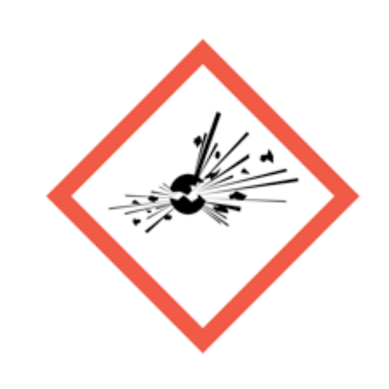
Exploding bomb
Explosive, self-reactive or organic peroxide hazards.
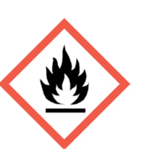
Flame
Flammable, self-heating or pyrophoric substances.
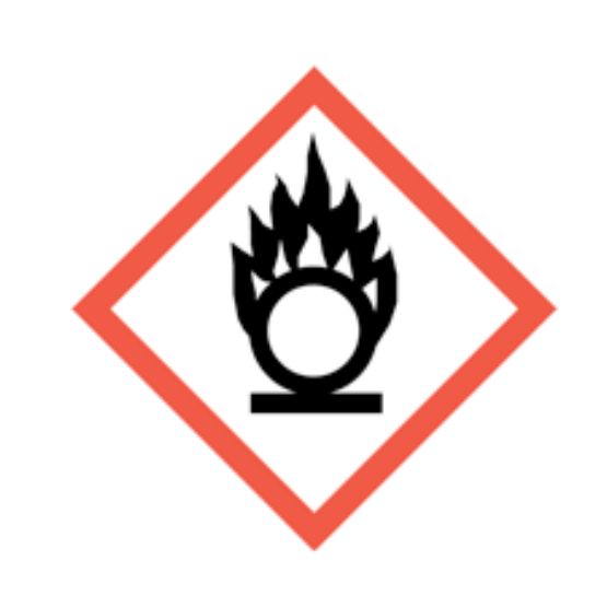
Flame over circle
Oxidising substances that can intensify fire.

Gas cylinder
Gas under pressure.
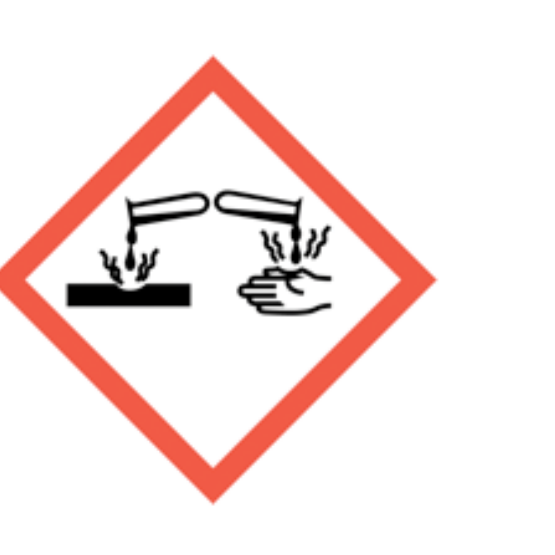
Corrosion
Skin burns, eye damage or corrosive to metals.
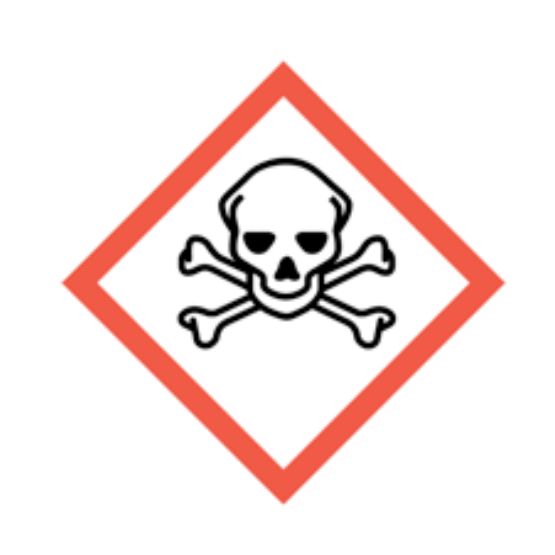
Skull and crossbones
Acute toxicity.
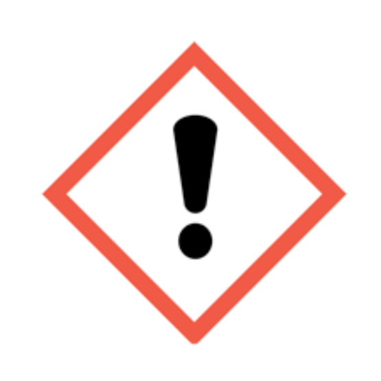
Exclamation mark
Irritation, harmful effects or skin sensitisation; some classifications cover drowsiness, dizziness or ozone-layer hazard. Read the actual hazard statements. Skin sensitisation differs from respiratory sensitisation.
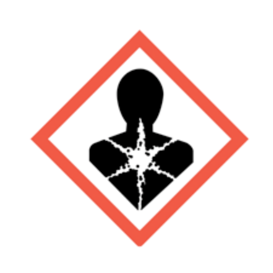
Health hazard
Serious health hazards, including organ damage, aspiration hazard, respiratory sensitisation, cancer and reproductive effects; some can follow a single exposure.
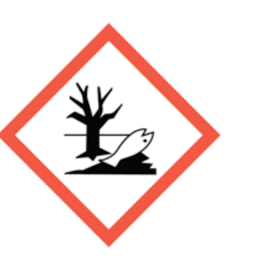
Environment
Hazardous to the aquatic environment.
What acronyms are used on this page?
Acronyms should be explained so chemical eye-hazard controls are accessible.
| Acronym | Meaning | Why it matters |
|---|---|---|
| CLP | Classification, Labelling and Packaging | Chemical classification system shown on labels and SDS. |
| COSHH | Control of Substances Hazardous to Health | Main GB framework for hazardous substances. |
| EH40 | HSE workplace exposure limits publication | Reference for workplace exposure limits. |
| PPE | Personal Protective Equipment | Includes goggles, face shields, gloves and protective clothing. |
| RPE | Respiratory Protective Equipment | May be relevant where vapours, dusts or aerosols also affect breathing. |
| SDS | Safety Data Sheet | Supplier information on hazards, controls and first aid. |