SHARE Knowledge Hub
Bursitis, including prepatellar bursitis
Bursitis is inflammation of a bursa, a small fluid-filled sac that cushions tissues around a joint and helps reduce friction between skin, tendons, muscles and bone. In workplace health, the classic example is prepatellar bursitis, often called housemaid's knee, where the bursa in front of the kneecap becomes swollen and painful after prolonged or repetitive kneeling, direct pressure, friction or trauma. Bursitis can affect other joints too, including the elbow, shoulder, hip and heel, but occupational controls are strongest where the exposure is clear: kneeling, pressure, awkward lower-level work, repeated contact stress, poor padding, hard surfaces and inadequate recovery. Good management means redesigning work to reduce pressure first, using knee protection and task rotation where needed, encouraging early reporting, protecting cuts from infection, and supporting workers through medical or occupational health advice when symptoms are severe, recurrent, infected or affecting safe work.
Understanding Bursitis, including prepatellar bursitis
Bursitis is inflammation of a bursa, a small fluid-filled sac that cushions tissues around a joint and helps reduce friction between skin, tendons, muscles and bone. In workplace health, the classic example is prepatellar bursitis, often called housemaid's knee, where the bursa in front of the kneecap becomes swollen and painful after prolonged or repetitive kneeling, direct pressure, friction or trauma. Bursitis can affect other joints too, including the elbow, shoulder, hip and heel, but occupational controls are strongest where the exposure is clear: kneeling, pressure, awkward lower-level work, repeated contact stress, poor padding, hard surfaces and inadequate recovery. Good management means redesigning work to reduce pressure first, using knee protection and task rotation where needed, encouraging early reporting, protecting cuts from infection, and supporting workers through medical or occupational health advice when symptoms are severe, recurrent, infected or affecting safe work.
Why this matters at work
A floor-laying or cleaning task may need several connected assessments: local contact pressure, load handling, awkward reach, skin injury, access and other hazards. Use the relevant tool only for the part it addresses and retain the observations and decision basis, not the score alone. Assess the revised complete method for new force, instability or awkward upper-limb work. Record remaining low tasks and practical support. A wider cleaning head or raised component may reduce one exposure while increasing another. Verify the actual task under representative conditions and obtain competent advice where the chosen tools do not cover the mechanism.
Watch for workarounds, repeated missing equipment, pad complaints, contact with debris, delayed reporting and task changes that recreate low-level work. A cluster warrants review of exposure and support, not an automatic claim of common occupational causation. Compare patterns with the actual work and reporting culture. A fall in reported discomfort may follow fear of consequences rather than a successful control. Avoid targets that reward managers for having no reports or pressure workers to return before suitable arrangements exist. Use indicators to ask a concrete question and allocate action. Keep the smallest useful dataset, explain how it is used and verify whether improvements persist after the initial campaign or equipment issue.
How organisations manage this in practice
Distinguish what has genuinely been eliminated from what has been moved elsewhere. Factory preparation can leave site finishing, inspection, adjustment or maintenance exposure. Record these remaining tasks and make responsibility for them clear. A design review should ask who will install, clean, inspect and repair the asset through its life. A worker's inability to suggest a different method is not proof that elimination is impracticable. Obtain design or engineering input where needed and keep a reasoned record of the decision. Demonstrate the new sequence with actual components before claiming the exposure has disappeared; the measurement is whether the relevant contact still occurs during the work, not whether the method has acquired a new name.
Investigate a control failure through actual conditions: access, equipment, supply, sequence, workload, supervision and the worker's account. A worker seen without a pad may have been issued one that slips or cannot be used for that task. Establish facts and fix the system while applying any fair organisational process separately. Do not let punitive assumptions discourage early reporting or replace an unresolved engineering choice. Record disagreements and uncertainty rather than writing a predetermined conclusion. Feed the findings back to those involved, including the action owner and verification route. Consultation is useful only when the organisation can respond to what people identify and show what changed.
A useful task description connects the contact to its source: low fixing points, floor seams, narrow access, hard or uneven surfaces and repeated transitions between kneeling and standing. Note whether the person supports body weight on one knee, slides along the floor or rests the elbow on a bench edge. Work can aggravate a condition even where it did not initiate it. Investigate exposure and control failures without requiring proof of exclusive occupational causation before making the task safer. Avoid an inevitable progression diagram: workers differ, and symptoms, examination and investigations govern the clinical interpretation.
Swelling, heat, redness, fever, feeling unwell, severe pain, inability to move a joint, or symptoms that are worsening should be assessed through appropriate medical routes. Managers should not diagnose bursitis; they should reduce aggravating work exposure, support early reporting, consider occupational health advice and review the task that is putting pressure on the joint.
Reduce knee contact without transferring the load
Review how much knee contact the task creates, the surface and the work sequence. Stronger changes can remove or reduce kneeling before residual protection is selected. A new tool must be checked for reach, back, grip and handling demands rather than judged only on knee contact.
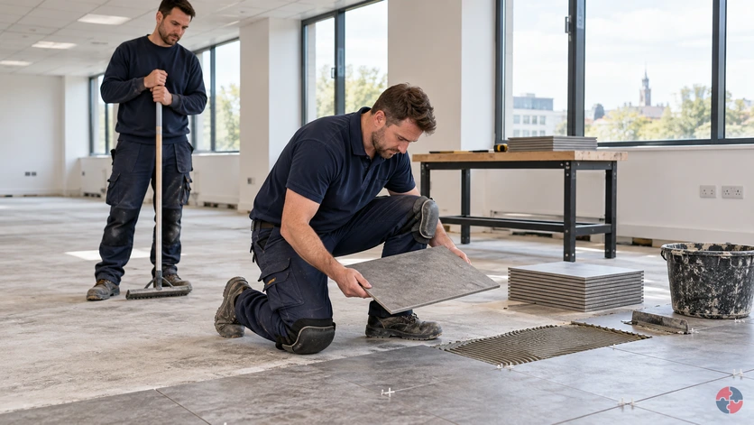
Review the floor task beyond the knee pads
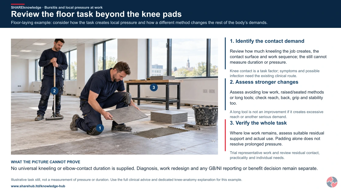
Full written explanation
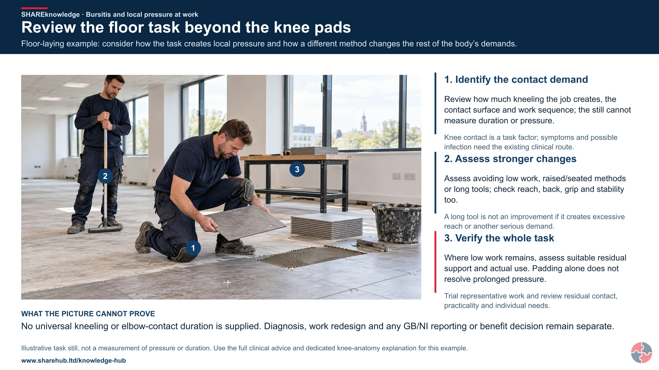
Floor-laying example: consider how the task creates local pressure and how a different method changes the rest of the body’s demands.
- Identify the contact demand. Review how much kneeling the job creates, the contact surface and work sequence; the still cannot measure duration or pressure.
- Assess stronger changes. Assess avoiding low work, raised/seated methods or long tools; check reach, back, grip and stability too.
- Verify the whole task. Where low work remains, assess suitable residual support and actual use. Padding alone does not resolve prolonged pressure.
Change the pathway
- No diagnosis from a picture. Knee contact is a task factor; symptoms and possible infection need the existing clinical route.
- Check transferred demands. A long tool is not an improvement if it creates excessive reach or another serious demand.
- Use worker feedback. Trial representative work and review residual contact, practicality and individual needs.
Evidence check: No universal kneeling or elbow-contact duration is supplied. Diagnosis, work redesign and any GB/NI reporting or benefit decision remain separate.
Full symptoms and appropriate clinical escalation context
Primary references: HSE: lower limb disorders · NHS: bursitis and when to obtain help
Open the complete HD image (new tab)Download image
Full symptoms and appropriate clinical escalation context
Primary references: HSE: lower limb disorders · NHS: bursitis and when to obtain help
Individual swelling and possible infection follow the existing clinical route; the supervisor does not diagnose. Keep diagnosis, GB/NI reporting and benefit eligibility separate. The operational redesign sheet needs task facts and control evidence, not personal clinical detail.
A raised-tool trial improves one demand but worsens another
Illustrative example. A floor layer trials a long-tool method that reduces kneeling but creates excessive reach at the far edge.
Decision. Reject the incomplete improvement and adapt the work height, tool or sequence until the overall task is better.
Evidence to check. Compare knee-contact baseline, surface, reach/back/handling effects, residual contact and worker feedback.
Hold or escalation point. Do not retain a method solely because kneeling time falls while another substantial demand increases.
Follow-up. Verify the selected method on representative floor areas and review residual protection and recovery arrangements.
Change the floor task before relying on pads
The prepatellar bursa lies in front of the kneecap. Compare methods by what they change in knee contact and in the rest of the task, rather than assume padding makes prolonged kneeling acceptable.
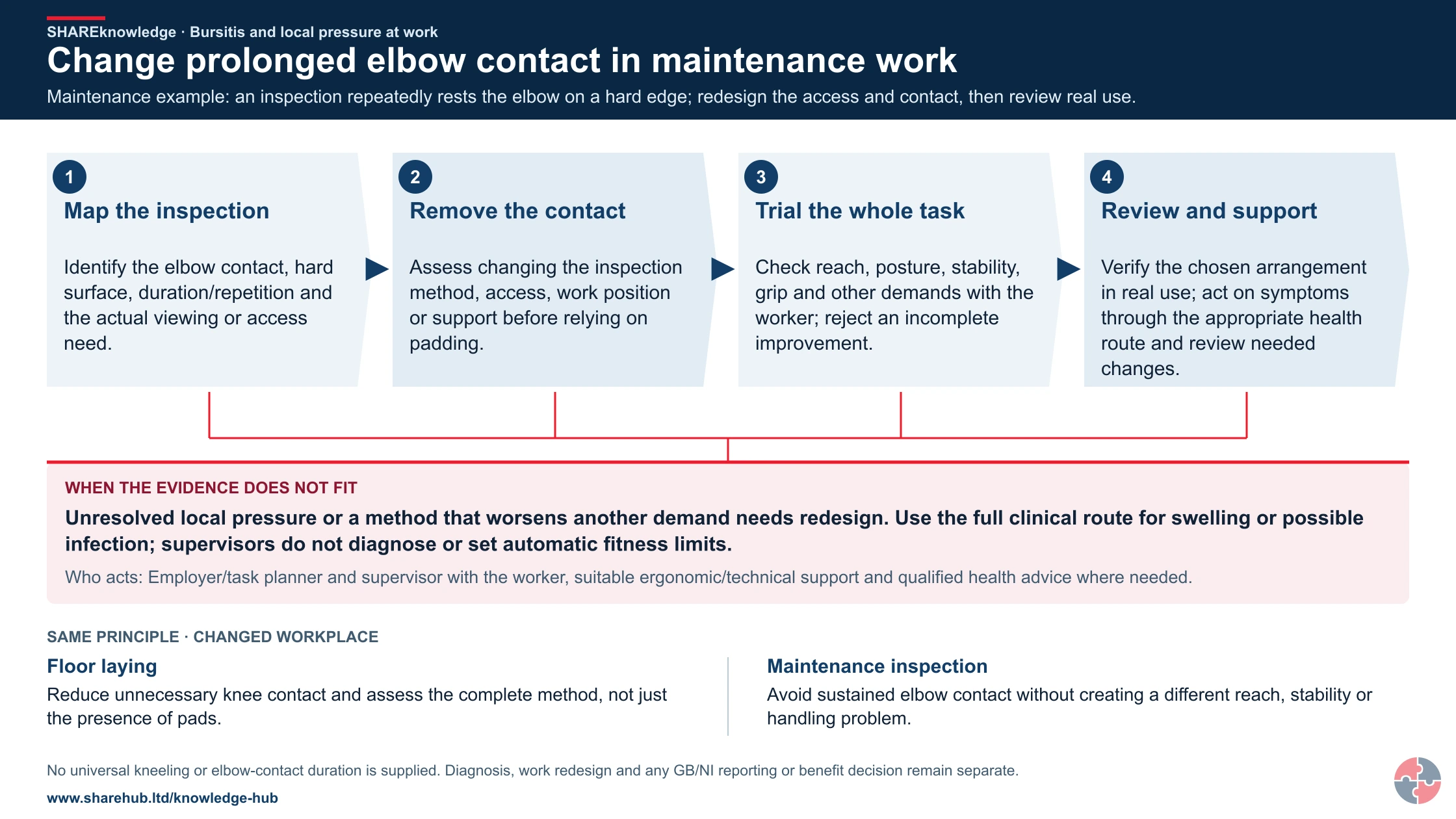
Change prolonged elbow contact in maintenance work
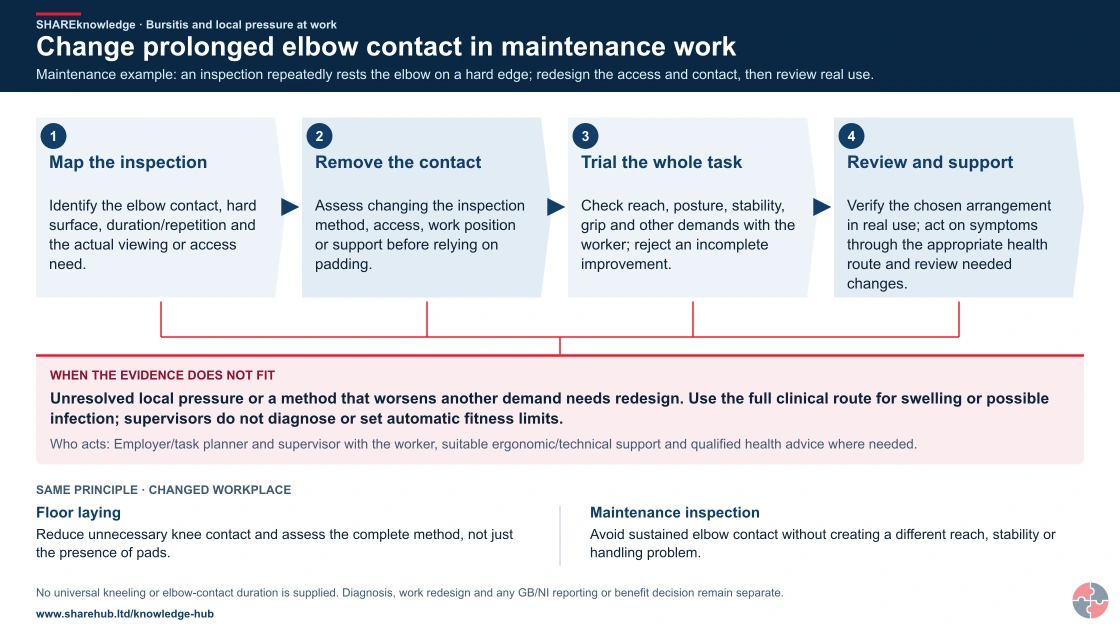
Full written explanation
Maintenance example: an inspection repeatedly rests the elbow on a hard edge; redesign the access and contact, then review real use.
- Map the inspection. Identify the elbow contact, hard surface, duration/repetition and the actual viewing or access need.
- Remove the contact. Assess changing the inspection method, access, work position or support before relying on padding.
- Trial the whole task. Check reach, posture, stability, grip and other demands with the worker; reject an incomplete improvement.
- Review and support. Verify the chosen arrangement in real use; act on symptoms through the appropriate health route and review needed changes.
When the evidence does not fit: Unresolved local pressure or a method that worsens another demand needs redesign. Use the full clinical route for swelling or possible infection; supervisors do not diagnose or set automatic fitness limits.
Who acts: Employer/task planner and supervisor with the worker, suitable ergonomic/technical support and qualified health advice where needed.
Changed workplace
Floor laying: Reduce unnecessary knee contact and assess the complete method, not just the presence of pads.
Maintenance inspection: Avoid sustained elbow contact without creating a different reach, stability or handling problem.
Evidence check: No universal kneeling or elbow-contact duration is supplied. Diagnosis, work redesign and any GB/NI reporting or benefit decision remain separate.
Full symptoms and appropriate clinical escalation context
Primary references: HSE: lower limb disorders · NHS: bursitis and when to obtain help
Open the complete HD image (new tab)Download image
Full symptoms and appropriate clinical escalation context
Primary references: HSE: lower limb disorders · NHS: bursitis and when to obtain help

- Remove unnecessary floor work
- Assess raised / seated method
- Residual support + whole-task check
Read the labels in the full-size drawing
- Avoid unnecessary floor
- assembly
- Assess raised / seated work
- Residual support + whole
- task
- Pressure, reach, back load, stability and handling all matter
| Option | Pressure question | Transferred-risk check |
|---|---|---|
| Remove unnecessary floor assembly | Can preassembly or a different sequence remove the need to kneel? | Handling the larger assembly, installation access and stability. |
| Suitable raised or seated method | Can the work be brought to an appropriate level or supported differently? | Reach, back loading, stable support and the actual fitting task. |
| Assessed residual low work | What suitable support is needed when low-level work remains? | Duration, repetition, movement, trips, hygiene and whether the task still needs redesign. |
| Worker review | Does the revised method work for the individual and the work? | Symptoms, practical feedback, adjustments and the qualified health route where needed. |
Use the dedicated knee inset for this example. Other regional anatomy is optional wider learning and must not substitute for it. No universal kneeling duration, treatment or automatic fitness decision is given.
What are the key facts about bursitis?
The key facts are that bursitis is local soft-tissue inflammation, often preventable where pressure and kneeling exposure can be reduced.
Bursae reduce friction
They sit around joints and allow tissues to glide; inflammation causes swelling, tenderness and pain.
Kneeling is a classic work exposure
HSE identifies knee bursitis as associated with repetitive kneeling.
Infection changes the risk
Heat, redness, fever, chills or feeling unwell require prompt medical assessment.
Padding helps but is not the first control
Knee pads reduce pressure but do not remove the need to redesign work and reduce kneeling time.
Early reporting supports exposure reduction
Report symptoms early so harmful knee pressure can be reduced and qualified advice sought; this page does not predict an individual recovery time or treatment outcome.
Return to the same task needs planning
If the exposure is unchanged, recurrence is more likely.
What is bursitis?
Bursitis is inflammation of a small cushioning sac around a joint. See DWP/IIAC occupational beat disorders report (July 2006)
A bursa is a small fluid-filled sac that reduces friction where skin, tendons, muscles or ligaments move over bone. When a bursa becomes irritated, it can produce excess fluid and become swollen, painful and tender.
Bursitis can affect several joints. In occupational health, the most relevant example is often prepatellar bursitis at the knee because the prepatellar bursa lies directly between the skin and kneecap, where it is exposed to pressure from kneeling.
The dated IIAC report distinguishes occupational inflammation or infection of a bursa. Pressure-related inflammation and possible infection require different clinical assessment; a skin break with a hot, red swelling is a reason to seek advice, not a workplace diagnosis. Nidirect’s MRSA information includes septic bursitis as one possible infection outcome, but neither this example nor those symptoms establish MRSA or any specific organism.
Bursa. A small cushioning sac around a joint. Can become irritated by pressure or friction.
Bursitis. Inflammation and swelling of a bursa. May affect kneeling, walking, bending or tool use.
Prepatellar bursitis. Bursitis in front of the kneecap. Classic occupational example linked to kneeling.
Aseptic bursitis. Inflammation without infection. Often linked to pressure, trauma or overuse.
Septic bursitis. Bursitis caused by infection. May follow cuts or skin breaks and needs medical assessment.
How does bursitis develop in the body?
Bursitis develops when pressure, friction, trauma or infection irritates the bursa and fluid builds up.
Pressure on the front of the knee can irritate the bursa


Read the complete visual as text
- NORMAL FUNCTION: The small prepatellar bursa lies in front of the kneecap and helps tissues move with less friction.
- WORK DEMAND: Prolonged or repeated kneeling, direct pressure and friction can irritate the bursa and cause swelling or pain.
- REDESIGN: Try raised or seated work and suitable longer tools first. Check reach, back loading, stability and other transferred risks.
- RESPONSE: Pads and shorter bouts may help residual contact but do not make painful kneeling acceptable. A hot or rapidly worsening knee needs prompt clinical assessment.
- Side-view knee schematic, not to scale: femur above tibia, kneecap (patella) in front of the joint, prepatellar bursa in front of the kneecap, and skin surface outside it.
- Repeated kneeling places pressure on the prepatellar bursa; the drawing is not diagnosis.
In prepatellar bursitis, the mechanism is easy to picture. The bursa sits just beneath the skin at the front of the knee. Repeated kneeling on a hard surface compresses the bursa between the floor and kneecap. Over time, the tissue becomes irritated, produces more fluid and swells.
Once swollen, the bursa becomes more vulnerable. Continuing to kneel directly on it can maintain the inflammation, increase tenderness and make movement or bending uncomfortable. A scrape or cut provides an entry risk for bacteria, so hot, red swelling with a skin break needs clinical advice; staff must not decide at work whether the bursa, skin or joint is infected.
The workplace control message is therefore straightforward: reduce direct pressure, reduce duration, improve surfaces, protect the skin, rotate tasks, provide recovery and act early.
Exposure. Kneeling, pressure, friction, direct blow or skin break. Identify pressure points and task duration.
Irritation. Bursa becomes inflamed and produces extra fluid. Reduce kneeling and direct contact early.
Swelling. Soft swelling forms over the joint. Modify tasks and avoid pressure on the swelling.
Functional impact. Pain, tenderness and reduced movement affect work. Consider temporary restrictions and OH advice.
Complication. Infection may occur if bacteria enter the bursa. Escalate hot, red, worsening or systemic symptoms.
The linked July 2006 IIAC report is used only for anatomy and occupational background: it describes the prepatellar bursa between skin and kneecap, inflammation, increased fluid and historical kneeling exposure. Current clinical assessment and current statutory reporting must use their separate sources.
How does a working hand move, and what changes in nerve and tendon problems?
Interactive anatomy · Tendons, nerves and cushioning tissues · Updated 2026-10-23
Read all anatomy and process explanations without the viewer
General anatomy and example mechanisms, not diagnosis, treatment choice or fitness-for-work assessment. These are examples of mechanisms. Similar symptoms can have different causes. Self-provocation or nerve-tapping tests Exercise or stretching prescriptions Automatic symptom classification or a generic RSI lesion Treatment, return-to-work clearance or a task-based diagnosis Unreviewed shoulder, medial-elbow or trigger-finger damage overlays The prepatellar-bursitis route requires the dedicated labelled front-of-knee inset. A wrist/shoulder substitute is not suitable. Prompt-help text remains outside the model and scenarios.
Forearm flexor muscles
These muscles contract and pull through tendons to bend the fingers and thumb. The forearm flexor muscles generate the pull transmitted through tendons into the fingers and thumb. As the illustrated grip closes, follow the muscle region towards those slender connections. Their action bends the finger joints; the tendon transfers the pull rather than generating it. The visible shape change makes this relationship clearer and does not measure muscle effort. Gloucestershire Hospitals NHS Foundation Trust: Flexor Tendon Injuries
Flexor tendons
These tendons carry muscle force into the hand. Flexor tendons connect forearm muscles with bones in the hand. Their glide lets a muscle’s pull bend the finger joints while guided paths keep the tendons near the skeleton. Nine flexor tendons pass through the carpal tunnel. Compare their mechanical role with the nearby median nerve, which carries signals rather than pulling on bone. Gloucestershire Hospitals NHS Foundation Trust: Flexor Tendon Injuries · Whittington Health NHS Trust: Carpal Tunnel Syndrome
Tendon sheath
The sheath surrounds the tendon and supports smooth glide. A tendon sheath surrounds and supports a tendon as it glides. Along the fingers, pulley structures help keep the tendon close to the bones so its pull acts along a guided route. Watch the tendon move relative to its surroundings. A sheath is part of that supporting passage, not another muscle producing the same pull. Gateshead Health NHS Foundation Trust: Trigger Finger/Thumb Release Surgery
Carpal bones
Eight small carpal bones make up the wrist. Their curved arrangement forms the bony side of the carpal tunnel. The transverse carpal ligament closes the tunnel across the palm side. King’s College Hospital NHS Foundation Trust: Carpal tunnel syndrome — patient information · Kent Community Health NHS Foundation Trust: Carpal tunnel syndrome guide for patients
Transverse carpal ligament
This strong ligament spans the palm side of the wrist. It forms the roof above the median nerve and flexor tendons. Separating layers in the model helps reveal these relationships. King’s College Hospital NHS Foundation Trust: Carpal tunnel syndrome — patient information · Whittington Health NHS Trust: Carpal Tunnel Syndrome
Median nerve
The median nerve carries signals for feeling and some thumb movement. The median nerve carries sensory information from the thumb, index and middle fingers and part of the ring finger, and helps control some thumb muscles. Follow its branches beyond the shared wrist passage. The cyan travel cue represents communication; it is not a visible electrical flash, a nerve-speed measurement or a map for diagnosing altered sensation. NHS: Carpal tunnel syndrome · Kent Community Health NHS Foundation Trust: Carpal tunnel syndrome guide for patients · Whittington Health NHS Trust: Carpal Tunnel Syndrome
Thumb tendon: APL
Abductor pollicis longus helps move the thumb away from the hand. APL passes along the thumb side of the wrist through the first dorsal compartment. Follow it towards the base of the thumb. Ashford and St Peter’s Hospitals NHS Foundation Trust: De Quervains · Wrightington, Wigan and Leigh Teaching Hospitals NHS Foundation Trust: De Quervain’s Syndrome
Thumb tendon: EPB
Extensor pollicis brevis helps straighten the thumb. EPB travels beside APL through the first dorsal compartment on the thumb side of the wrist. Ashford and St Peter’s Hospitals NHS Foundation Trust: De Quervains · Wrightington, Wigan and Leigh Teaching Hospitals NHS Foundation Trust: De Quervain’s Syndrome
First dorsal compartment
A small passage on the thumb side of the wrist guides APL and EPB. The tendons normally glide through their sheath here. This is the location of the De Quervain’s example; the carpal tunnel lies on the palm side. Ashford and St Peter’s Hospitals NHS Foundation Trust: De Quervains · Wrightington, Wigan and Leigh Teaching Hospitals NHS Foundation Trust: De Quervain’s Syndrome
Outer elbow tendon attachment
Forearm tendons attach to bone near the outside of the elbow. Forearm extensor tendons attach near the bony point on the outside of the elbow, the lateral epicondyle. These muscles extend and help steady the wrist during gripping. Link the closer attachment view with the tool task to see why an elbow structure matters during hand use. Pain here does not establish a torn or detached tendon. NHS: Tennis elbow · Berkshire Healthcare NHS Foundation Trust: Advice and resources: Lateral elbow tendinopathy · Newcastle Hospitals NHS Foundation Trust: Tennis elbow
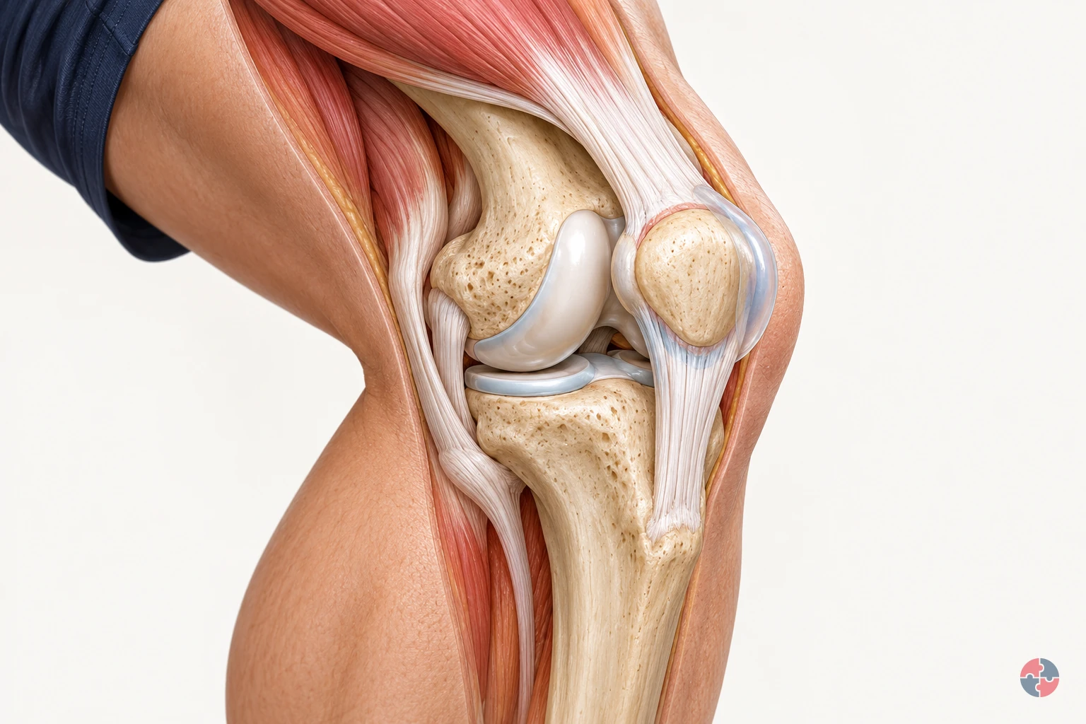
Prepatellar bursa
A fluid-containing sac helps reduce friction at the front of the kneecap. The prepatellar bursa lies between the kneecap and the overlying soft tissues, outside the knee joint. Its fluid-containing structure helps cushioning and smooth glide over bone. Compare the closer view with the worker’s knee contact in the floor task. The normal view locates the sac; it does not diagnose inflammation or measure pressure during kneeling. NHS: Bursitis · Leeds Community Healthcare NHS Trust: About pre-patella bursitis
Open the complete process transcript
The process, step by step
Watch muscles pull and tendons glide
The forearm muscles provide much of the pull that closes the fingers around an object. When these muscles contract, their tendons transmit force into the hand and pull on the finger bones, bending the joints. The tendons glide along guided paths rather than pulling away from the fingers like loose cords. Follow the muscle-to-tendon connection as the illustrated grip closes, then follow the return movement as the hand opens. The model enlarges this motion so that you can connect the movement at the fingers with the working structures farther up the forearm. Gloucestershire Hospitals NHS Foundation Trust: Flexor Tendon Injuries · Gateshead Health NHS Foundation Trust: Trigger Finger/Thumb Release Surgery
What to watch: Look from the forearm muscle bellies towards the slender flexor tendons, then follow their paths into the fingers around the handle.
Why it matters: Tendons transfer a muscle’s mechanical pull to bone, allowing joints to move. Guided tendon paths help direct that pull through the hand; the nerve has a different role in signalling.
Explore this step: Pause during the grip and trace one tendon from its forearm muscle towards the finger.
Watch tendon glide through the wrist
The carpal tunnel is a passage at the wrist, with a bony boundary formed by the carpal bones and a strong ligament spanning the palm side. Nine flexor tendons and the median nerve pass through this shared space. The tendons transmit the pull that helps bend the fingers and thumb; the nerve carries signals. Follow the tendon movement beneath the ligament roof and compare it with the nerve’s position. The cutaway separates the layers for learning, but these structures normally remain close together within a connected wrist. King’s College Hospital NHS Foundation Trust: Carpal tunnel syndrome — patient information · Kent Community Health NHS Foundation Trust: Carpal tunnel syndrome guide for patients · Whittington Health NHS Trust: Carpal Tunnel Syndrome
What to watch: Find the carpal bones and ligament roof, then compare the flexor tendons with the median nerve passing through the same wrist passage.
Why it matters: The tunnel brings several structures through a compact part of the wrist. Understanding their different functions explains why tendon glide and nerve signalling must be explored separately even though they occupy nearby space.
Explore this step: Use the labels to identify the tunnel boundary, a tendon and the nerve without changing to an injury example.
Follow the nerve signal
The median nerve links part of the hand with the nervous system. It carries sensory information from the thumb, index finger, middle finger and part of the ring finger, and helps control some muscles at the base of the thumb. Follow the cyan travelling cue along the nerve and into its branches. This colour illustrates signalling; a real nerve does not visibly light up. Compare that pathway with the tendons beside it: tendons transfer mechanical pull, whereas the nerve carries information. The simplified branches are a teaching map, not a test of a person’s sensation. Kent Community Health NHS Foundation Trust: Carpal tunnel syndrome guide for patients · Whittington Health NHS Trust: Carpal Tunnel Syndrome
What to watch: Follow the cyan cue along the median nerve through the wrist into the palm, keeping the adjacent tendons in view.
Why it matters: Sensation helps the nervous system receive information from the hand, while motor signals contribute to thumb control. Distinguishing information from mechanical pull makes the coordinated hand function easier to understand.
Explore this step: Compare the nerve cue with tendon glide and explain which one represents information and which represents force transfer.
Follow the thumb tendons
Two tendons run along the thumb side of the wrist through the first dorsal compartment. They connect forearm muscles to the thumb and glide as the thumb changes position. Abductor pollicis longus, labelled APL, helps move the thumb away from the hand. Extensor pollicis brevis, labelled EPB, helps straighten it. Follow the two labelled paths rather than assuming they pass through the palm-side carpal tunnel. The model brings their movement and location into view so that the separate actions and their shared guiding passage can be understood together. Ashford and St Peter’s Hospitals NHS Foundation Trust: De Quervains · Wrightington, Wigan and Leigh Teaching Hospitals NHS Foundation Trust: De Quervain’s Syndrome
What to watch: Find APL and EPB beside the thumb side of the wrist, then follow both labelled paths through the first dorsal compartment.
Why it matters: Different tendon actions contribute to positioning the thumb for hand use. Their shared passage guides glide, but the two tendons retain different attachments and functions; they are not interchangeable parts.
Explore this step: Select each tendon in turn and connect its name with the thumb movement described in the explanation.
Follow the elbow during a tool task
A tool task involves more than the muscles that visibly close the fingers. Forearm extensor muscles also help hold the wrist steady while the hand grips. Follow these muscles back towards their tendon attachment near the bony point on the outside of the elbow, called the lateral epicondyle. Watch the technician’s illustrated grip and tool turn, then use the closer elbow view to connect the activity with this attachment. A moving tool does not demonstrate a damaged tendon: the normal task and the separate tissue example explain different things. NHS: Tennis elbow · Newcastle Hospitals NHS Foundation Trust: Tennis elbow
What to watch: Watch the hand grip the screwdriver, then follow the working forearm towards the tendon attachment on the outside of the elbow.
Why it matters: Wrist stability helps the hand grip and use an object, so a tendon attachment near the elbow can be involved in a hand task. Movement alone does not show injury or quantify the demand.
Explore this step: Switch between Work activity and Elbow cutaway and describe how the two views relate to the same task.
Locate the bursa in front of the kneecap
The prepatellar bursa is a small fluid-containing sac in front of the kneecap. It helps the overlying soft tissues move across the bone with less friction. First locate the front of the knee in the floor-laying task, then follow the linked closer view to the thin sac outside the knee joint. This shows where contact during kneeling relates to the anatomy; the bursa is not a cushion inside the joint. The normal location lesson remains separate from the example of bursal irritation, and the short model sequence does not predict when symptoms would occur. Leeds Community Healthcare NHS Trust: About pre-patella bursitis · NHS Musculoskeletal Matters Dorset: Prepatellar bursitis
What to watch: Locate the kneeling worker’s front-of-knee contact, then find the thin bursa between the kneecap and the overlying soft tissues in the close view.
Why it matters: Glide between soft tissue and bone is a different function from movement inside the knee joint. Locating the bursa makes the relevant contact area clear without inferring tissue damage from posture alone.
Explore this step: Compare Floor-laying task with Knee cutaway and identify which side of the kneecap contains the bursa.
Mechanisms and their limits
Prepatellar bursitis: front-of-knee cushion Repeated or prolonged kneeling can irritate the small bursa in front of the kneecap. It may become inflamed and swollen; direct injury or infection can also cause bursitis. This example highlights the prepatellar bursa in front of the kneecap, with the knee joint kept distinct behind it. The enlarged bursa and local colour are teaching cues. Symptoms vary; the model does not predict swelling or measure its severity. A hot swollen knee with fever or feeling unwell needs urgent professional advice; do not assume it is just work-related irritation. NHS: Bursitis · Leeds Community Healthcare NHS Trust: About pre-patella bursitis · NHS Musculoskeletal Matters Dorset: Prepatellar bursitis · Health and Safety Executive: Lower limb disorders
NHS: prepatellar bursitis and kneeling
HSE: reduce kneeling and lower-limb risks
Prepatellar bursitis: front-of-knee cushion — follow the anatomy
Look at: The small fluid-filled bursa in front of the kneecap.
What changes: Prolonged pressure from kneeling can irritate this cushioning sac.
Why it matters: Swelling and tenderness can develop; infection and other causes need clinical assessment.
What to do: Reduce kneeling time, use suitable support and seek advice for marked, hot or worsening swelling.
Prolonged kneeling while laying flooring
A floor layer kneels on both knees and moves a hand float across a tiled area, repeating the work close to the floor. A teaching time jump separates the kneeling task from a possible bursa-swelling example. Playback speed and time do not set a safe kneeling limit or predict when symptoms develop. Kneeling does not inevitably cause bursitis, and padding does not instantly heal an affected bursa. Infection and direct injury are other possible causes. A hot, swollen knee with fever or feeling unwell needs urgent advice.
Watch the floor layer at work Watch the floor layer kneel on both knees and move the hand float across the tiled area. Notice where the knees meet the floor and how repeated reaches keep pressure on the front of the knees. Leeds Community Healthcare NHS Trust: About pre-patella bursitis · NHS Musculoskeletal Matters Dorset: Prepatellar bursitis · Health and Safety Executive: Lower limb disorders
Follow contact pressure at the knee Follow the contact cue from the floor towards the front of the kneecap. Repeated or prolonged kneeling can irritate the small prepatellar bursa here. The cue shows location, not a measured pressure. Leeds Community Healthcare NHS Trust: About pre-patella bursitis · NHS Musculoskeletal Matters Dorset: Prepatellar bursitis · Health and Safety Executive: Lower limb disorders
Compare a possible local bursa change The closer view shows a modestly enlarged bursa in front of the kneecap. Inflammation here can cause local swelling, tenderness or pain. The coloured area is a teaching cue; symptoms vary and this example cannot diagnose a knee. NHS: Bursitis · Leeds Community Healthcare NHS Trust: About pre-patella bursitis
Reduce contact pressure and kneeling time Compare the cushion beneath both knees and the closer, shorter working strokes. Change methods and vary tasks to reduce time spent kneeling. Suitable padding can reduce contact pressure, but persistent symptoms and warning signs still need appropriate advice. Leeds Community Healthcare NHS Trust: About pre-patella bursitis · NHS Musculoskeletal Matters Dorset: Prepatellar bursitis · Health and Safety Executive: Lower limb disorders
Read the practice situations and all answer explanations
UL-Q1: Which change should lead the review?
A packer repeats a forceful grip for long periods.
Only teach stretching. — Stretching alone leaves the work demands in place. Review the task, tools, layout and organisation with workers.
Review force, repetition, workstation fit, task duration and recovery opportunities. — Yes. Address the demands creating risk and check that changes work in practice. Early symptom reporting and suitable occupational-health advice also matter.
Use a wrist-posture score to diagnose the condition. — An ergonomic assessment helps identify work risks. It does not diagnose a medical condition.
Health and Safety Executive: Upper limb disordersUL-Q2: Which structure is central to the carpal-tunnel card?
Compare the three labelled locations in this teaching model.
The median nerve under pressure at the wrist. — Correct. Select UL-06 in the front-of-wrist view. It is a nerve-compression example.
The thumb-side sheath containing APL and EPB. — That is the thumb-side first dorsal compartment: UL-07 to UL-09 in the De Quervain’s view, not the carpal tunnel.
A swollen bursa in front of the kneecap. — That is UL-11 in the dedicated knee inset, relevant to prepatellar bursitis.
NHS: Carpal tunnel syndrome · Ashford and St Peter’s Hospitals NHS Foundation Trust: De Quervains · NHS: Bursitis · Leeds Community Healthcare NHS Trust: About pre-patella bursitisUL-Q3: What can this model tell them?
A learner notices pain at the outside of their elbow.
The animation proves a tendon has ruptured. — The model does not establish rupture or show a person’s tissues. Sudden severe injury or loss of function needs appropriate clinical advice.
This site may be relevant to lateral elbow tendinopathy, but an image cannot diagnose the person. — Correct. The location helps explain one example. Similar symptoms have different causes, and persistent or worsening symptoms deserve clinical advice.
All upper-limb pain is carpal tunnel syndrome. — Different nerves, tendons, joints and other structures can cause symptoms. The carpal-tunnel example is only one mechanism.
NHS: Tennis elbow · NHS: Tendonitis · Berkshire Healthcare NHS Foundation Trust: Advice and resources: Lateral elbow tendinopathySources and how the model is made
The opening images are AI-generated anatomical illustrations. The interactive 3D models and labelled SVG diagrams use original procedural artwork. These teaching images are not medical scans or diagnostic tools. Source references support the learner explanations and do not grant artwork rights.
- Health and Safety Executive: Upper limb disorders — Multiple work factors, task redesign, force/repetition/posture/duration, worker involvement and early reporting. Risk scores are not diagnosis.
- NHS: Carpal tunnel syndrome — Median-nerve pressure, potential sensory and functional effects, varied contributors and clinical assessment. No treatment or self-test generated.
- King’s College Hospital NHS Foundation Trust: Carpal tunnel syndrome — patient information — Bony tunnel boundary and transverse carpal-ligament roof. Text only, no third-party credited artwork copied. Printed review date September 2026 requires recheck at clinical sign-off; corroborates NHS anatomy.
- Ashford and St Peter’s Hospitals NHS Foundation Trust: De Quervains — APL and EPB actions and thumb-side first dorsal compartment. Indexed primary-page text rechecked, updated 28 April 2026; corroborated by directly retrieved UL8. No treatment recommendations or artwork copied.
- NHS: Tennis elbow — Outer-elbow location, tendon attachment and possible repeated gripping/twisting context. More precise tendinopathy wording corroborated by UL12/UL14; no blanket inflammation claim.
- NHS: Bursitis — Fluid-sac cushioning, inflammation and pressure-reduction context. Printed next-review date 30 October 2026 should be rechecked at sign-off.
- NHS: Tendonitis — General tendon role and urgent professional advice for severe pain or suspected rupture. Printed next-review date June 2026 is overdue: recheck before clinical approval. No treatment/exercise regimen reused.
- Wrightington, Wigan and Leigh Teaching Hospitals NHS Foundation Trust: De Quervain’s Syndrome — Directly retrieved January 2026 leaflet, expiry January 2028: APL/EPB, thumb-side first dorsal compartment and constrained sheath glide. Anatomy/mechanism only; no exercises or artwork copied.
- Gloucestershire Hospitals NHS Foundation Trust: Flexor Tendon Injuries — Directly retrieved: flexor tendons connect forearm muscles to finger bones and transmit their pull. Used for normal function only; no injury prognosis, treatment instructions or artwork reused.
- Gateshead Health NHS Foundation Trust: Trigger Finger/Thumb Release Surgery — Directly retrieved normal tendon/sheath distinction and guiding finger pulleys only. Printed review date September 2026 remains overdue; no trigger-finger overlay, surgical advice or exercise regimen reused.
- Ashford and St Peter’s Hospitals NHS Foundation Trust: Carpal Tunnel — Median-nerve sensory/motor role and flexor tendons within the tunnel. Indexed primary text verified, updated 28 April 2026; direct retrieval failed. Recheck live source at clinical sign-off.
- Berkshire Healthcare NHS Foundation Trust: Advice and resources: Lateral elbow tendinopathy — Painful forearm tendon attachment near the outside of the elbow and possible repetitive work demands. No treatment regimen, recovery prediction or artwork copied.
- Leeds Community Healthcare NHS Trust: About pre-patella bursitis — Dedicated bursa in front of kneecap, cushioning/glide, kneeling/direct injury/infection and urgent GP assessment for fever or feeling unwell. No treatment selection.
- NHS MyHealth Devon: Golfers & Tennis Elbow — Forearm tendon attachments and tendinopathy wording: not equated with simple inflammation. No rehabilitation or prognosis claims reused.
- NHS Musculoskeletal Matters Dorset: Prepatellar bursitis — Front-of-knee friction-reducing sac; prolonged kneeling, direct injury and infection as distinct possible causes. No medicine or cooling regimen reused.
- Kent Community Health NHS Foundation Trust: Carpal tunnel syndrome guide for patients — Directly retrieved, last edited 9 July 2025: eight carpal bones, ligament boundary, median-nerve sensory/motor role and tendon passage. Anatomy facts paraphrased only; no clinical regimen or artwork copied.
- Whittington Health NHS Trust: Carpal Tunnel Syndrome — Directly retrieved, updated 7 August 2025: nine tendons and median nerve in the carpal tunnel, nerve function and cautious attribution of work contribution. Normal anatomy corroborated by Sherwood Forest Hospitals NHS leaflet PIL202409-06-CTS; that leaflet has a September 2026 review date. No treatment recommendations or artwork reused.
- Newcastle Hospitals NHS Foundation Trust: Tennis elbow — Directly retrieved, last updated 3 November 2025: forearm tendons attach at the lateral epicondyle; these muscles extend the wrist and take part in gripping. Used for extensor anatomy and task explanation only; no treatment regimen, work-clearance advice or artwork copied.
- Health and Safety Executive: Lower limb disorders — Directly retrieved, updated 27 March 2025: repeated kneeling and knee bursitis; review working methods, mechanical aids, task variation, breaks, seating where possible, and suitable knee pads or matting. Used for prevention principles only; no numerical kneeling threshold, diagnosis or guarantee of protection.
What work factors can cause or aggravate bursitis?
Work factors include prolonged kneeling, repetitive kneeling, direct joint pressure, impact, poor surfaces and insufficient recovery. See HSE lower limb disorders
The strongest occupational pattern is pressure. Carpet fitting, floor laying, roofing, tiling, concrete finishing, maintenance, cleaning, gardening and some installation tasks can keep workers at floor level for long periods. If the worker kneels repeatedly on hard surfaces without suitable protection or breaks, the prepatellar bursa is repeatedly compressed.
Bursitis can also follow a direct blow or fall onto the knee, especially where the worker then continues kneeling. Infection risk increases where the skin is cut, grazed or punctured, particularly in dirty or contaminated environments.
Not every case is caused by work. Gout, rheumatoid arthritis, infection, sport, home activity and individual health factors can contribute. Employers still need to control work factors that could cause or worsen symptoms.
Prolonged kneeling. Compresses the prepatellar bursa for long periods. Redesign task, use platforms/seating, rotate work and limit duration.
Repetitive kneel-stand cycles. Adds friction and repeated loading. Plan task sequence and provide microbreaks.
Hard surfaces. Increase contact pressure at the knee. Use suitable padding, mats or alternative access.
Direct impact. Can inflame the bursa suddenly. Improve housekeeping, fall prevention and knee protection.
Skin cuts. Can allow infection to enter. Protect skin, clean cuts and use suitable first aid.
Poor work organisation. Keeps one worker on high-pressure tasks too long. Job rotation and realistic programme planning.
What symptoms can bursitis cause?
Bursitis commonly causes pain, swelling, tenderness, warmth and discomfort when moving or pressing on the joint. See nidirect knee pain and urgent help
In prepatellar bursitis, the swelling is usually visible or feelable at the front of the kneecap. The knee may feel tender when kneeling, bending, climbing or walking. Some people can move fairly well but cannot tolerate pressure on the front of the knee.
Symptoms that suggest possible infection are more concerning: increasing heat, redness, fever, chills, feeling unwell or rapidly worsening pain. A manager does not need to decide whether it is infected; the correct workplace response is to stop aggravating exposure and signpost to medical assessment.
Symptoms should be recorded as work impact, not diagnosis. Useful information includes which tasks are difficult, what exposure worsens symptoms, whether kneeling is required, whether the worker can move safely, and what temporary controls are needed.
Early irritation. Mild tenderness or swelling after kneeling. Reduce exposure and review task controls.
Established bursitis. Visible swelling, pain with pressure and reduced kneeling tolerance. Modify duties and consider OH/medical signposting.
Traumatic flare. Swelling after blow or fall. Investigate incident and reduce pressure.
Possible infection. Hot, red, swollen joint with fever, chills or feeling unwell. Stop exposure and use medical escalation route.
Recurrent bursitis. Repeated flare-ups after similar tasks. Redesign work, review PPE and improve job rotation.
How can bursitis affect work and future health?
Bursitis can limit kneeling, walking, bending, climbing, manual work and safe movement around the workplace.
For workers whose role depends on kneeling, even a small swelling can become a significant functional limitation. They may struggle to install flooring, fit skirting, work in roof spaces, service low-level plant, clean floors, garden, inspect low-level equipment or climb repeatedly.
Early reporting lets the employer reduce harmful pressure and arrange appropriate clinical advice. Returning to unchanged kneeling leaves the exposure in place; verify task redesign, residual protection and breaks rather than predict the worker’s prognosis.
Severe or infected bursitis can lead to longer absence, medical procedures and delayed return. This is why early reporting and sensible task modification should be normalised.
Kneeling intolerance. Worker cannot kneel comfortably. Avoid kneeling and use alternative access or task method.
Reduced mobility. Pain with bending, walking or climbing. Review access, travel distance and manual handling.
Manual task restriction. Difficulty working at floor level. Temporary task modification and rotation.
Skin vulnerability. Cuts or grazes around swollen knee. Skin protection, hygiene and first aid.
Absence or recurrence. Repeated flare-ups after same work. Root-cause review and ergonomic redesign.
How should work be controlled to prevent bursitis?
Controls should reduce kneeling pressure, duration, frequency and skin injury before relying on knee pads alone. See HSE lower limb disorders
The best control is to avoid or reduce kneeling. This might mean changing the work sequence, using adjustable benches, raising materials, using long-handled tools, prefabricating sections, using platforms, changing access, using mechanical aids or designing low-level work out during planning.
Where kneeling cannot be avoided, reduce exposure time. Rotate tasks, build in breaks, avoid keeping the same worker on floor-level tasks for the whole shift, and plan recovery after high-pressure work. Padding should be suitable for the surface, task and worker, and should not create new slip, trip or posture risks.
Controls should be checked at the workface. Knee pads left in the van, mats too small for the task, unrealistic programme pressure or workers kneeling outside the protected area all mean the control is not effective.
Task redesign. Raise work, use benches or use prefabrication. Has kneeling been reduced or removed?
Alternative tools. Use long-handled tools or extension equipment. Does the tool avoid floor-level pressure without creating awkward posture?
Work rotation. Share floor-level tasks and vary posture. Is exposure time genuinely reduced?
Breaks and recovery. Schedule breaks after kneeling tasks. Are breaks taken and protected?
Knee protection. Use suitable knee pads or mats. Are they comfortable, maintained and used correctly?
Skin care. Clean and cover cuts, maintain hygiene. Are cuts reported and protected?
Early reporting. Encourage reporting of swelling and pain. Are reports acted on before recurrence?
Where does kneeling remain after the main task is redesigned?
Observe preparation and finishing as well as the main operation. A standing method can reduce the longest kneeling period while floor-level checks, tool adjustments and clearing obstructions still require repeated knee contact. List those remaining activities and ask whether access, fixture height, tool design or the sequence can remove that contact too. Do not treat a reduction in the main task as proof that the whole shift is controlled.
For an illustrative flooring job using a standing tool, watch workers set up, reach edges, check the finish and resolve a fault. Agree which steps can be redesigned and which residual contact remains, then select any knee protection for the actual surface, fit, movement and other PPE. Check that the protection stays usable and that the revised method does not add awkward reach, force or instability. If a fault repeatedly restores prolonged kneeling, pause that method and correct the underlying access or tool problem. New or worsening symptoms still need the appropriate healthcare advice; a control trial cannot diagnose bursitis or override clinical restrictions.
Guidance and scope: Original SHARE whole-job example applying HSE GB and HSENI NI lower-limb guidance. No kneeling-duration threshold, pad certification requirement or promise of prevention or recovery is supplied. HSE lower limb disorders HSENI lower limb disorders
How can ERICPD be applied to bursitis prevention?
ERICPD helps prioritise task redesign and exposure reduction before relying on padding or personal discipline. See HSE lower limb disorders
Bursitis prevention is a good example of why the hierarchy of control matters. Knee pads are useful, but they still require the worker to kneel. Better controls remove or reduce the need to kneel, reduce time at floor level, reduce pressure, or change the method so the bursa is not repeatedly compressed.
PPE and discipline are still needed for residual risk: workers need comfortable knee protection, skin protection and the confidence to stop or rotate when symptoms appear. But those controls are weaker if the job is still designed around all-day kneeling.
Eliminate. Remove kneeling from the task. Use raised workstations or prefabricate at bench height.
Reduce. Reduce duration, frequency or pressure. Limit kneeling periods and use job rotation.
Isolate. Separate knees from hard surfaces. Use kneeling platforms or padded work surfaces.
Control. Plan work, breaks, task sequence and reporting. MSD risk assessment and supervisor checks.
PPE. Provide suitable knee pads or mats. Task-appropriate knee protection with maintenance.
Discipline. Train workers to report early and use controls. Toolbox talks, observations and feedback.
When should medical or occupational health support be considered?
Support should be considered where symptoms are severe, recurrent, worsening, possibly infected or affecting safe work. See nidirect knee pain and urgent help
Use an appropriate local clinical route for persistent or worsening knee symptoms or loss of movement. In Northern Ireland, nidirect advises immediate emergency-department assessment for a very painful knee or sudden pain, a badly swollen or changed-shape knee, or a very high temperature with feeling hot and shivery plus redness or heat around the knee. Do not delay that route for an occupational-health appointment; clinicians identify the cause.
Occupational health is useful where the condition affects work capability. The referral question should focus on function: what tasks should be avoided, how long restrictions may be needed, whether phased return or adjustments are appropriate, and whether further clinical advice is recommended.
Managers should avoid asking for unnecessary medical detail. The practical workplace need is to understand restrictions, controls, review dates and whether the task risk assessment needs changing.
Mild early symptoms. Self-care and early reporting, with medical advice if needed. Reduce kneeling and monitor.
Persistent symptoms. GP, physiotherapy or musculoskeletal service. Adjust duties and review task design.
Possible infection. Prompt medical assessment route. Stop exposure and protect the worker from further pressure.
Work capability affected. Occupational health advice. Agree restrictions, adjustments and review date.
Recurrent cases in team. Ergonomics or H&S review. Investigate task design and exposure patterns.
Do UK differences matter for bursitis reporting and support?
GB and NI reporting rules differ: current GB RIDDOR does not name knee bursitis, while NI Schedule 3 can require a beat-knee disease report when its diagnosis and work conditions are met. See NI RIDDOR Schedule 3
The prevention message is consistent across the UK: assess kneeling and pressure tasks, reduce exposure, provide suitable controls, encourage early reporting and support affected workers. However, reporting and compensation routes should not be assumed to be identical.
Current GB HSE RIDDOR occupational disease guidance does not list knee bursitis among reportable occupational diseases. GOV.UK Industrial Injuries Disablement Benefit technical guidance does list prescribed diseases A6 and A7 for bursitis or subcutaneous cellulitis at or about the knee or elbow due to severe or prolonged external friction or pressure. HSENI guidance for Northern Ireland identifies knee bursitis in its lower limb disorder guidance and references Northern Ireland RIDDOR requirements. See Government Industrial Injuries Disablement Benefit guidance See NI RIDDOR 1997 Schedule 3 See GB RIDDOR occupational diseases
For live cases, organisations should check the current official reporting route for the jurisdiction and avoid making assumptions from older guidance.
Great Britain RIDDOR. Current HSE occupational disease list. Knee bursitis is not listed in the current GB HSE page checked for this guide.
Industrial Injuries Disablement Benefit. GOV.UK prescribed disease list. A6/A7 include bursitis/subcutaneous cellulitis at knee or elbow due to severe or prolonged pressure/friction.
Northern Ireland. HSENI lower limb disorder and NI RIDDOR guidance. HSENI references reportability for knee bursitis under NI arrangements.
Occupational health support. Worker functional impact and adjustments. Support can be appropriate regardless of reportability.
Which legal duties apply to bursitis and kneeling work?
The legal and disease-reporting route depends on whether the work is in Great Britain or Northern Ireland; do not transfer a GB disease list to NI.
Employers must assess and control work that can cause or worsen musculoskeletal disorders. For bursitis, that means looking at kneeling, squatting, floor-level work, awkward posture, manual handling, repetitive pressure and whether controls are suitable for the real task.
Personal health information should be handled confidentially. Where symptoms persist, discuss practical adjustments individually and seek jurisdiction-specific HR or legal advice on any disability duty.
| Direct instrument or regulator route | Jurisdiction | How it applies | Practical evidence |
|---|---|---|---|
| Health and Safety at Work etc. Act 1974 | Great Britain | General GB duty to prevent work-related ill health. | Task and control review. |
| Management of Health and Safety at Work Regulations 1999 | Great Britain | GB assessment, arrangements and competent support. | Risk decision and action owner. |
| Health and Safety at Work (Northern Ireland) Order 1978 | Northern Ireland | Separate NI general duty. | NI task and control record. |
| Management of Health and Safety at Work Regulations (Northern Ireland) 2000 | Northern Ireland | Separate NI assessment and preventive arrangements. | NI risk decision. |
| Manual Handling Operations Regulations 1992 | Great Britain | Only where the kneeling task includes hazardous manual handling. | Handling assessment and redesign. |
| Manual Handling Operations Regulations (Northern Ireland) 1992 | Northern Ireland | NI manual-handling rule where the task includes hazardous handling. | NI handling assessment. |
| Personal Protective Equipment at Work Regulations 1992 | Great Britain | Suitable pads for residual knee-contact risk after stronger controls. | PPE selection and review. |
| Personal Protective Equipment at Work Regulations (Northern Ireland) 1993 | Northern Ireland | NI PPE route for residual knee-contact risk. | NI PPE selection. |
| GB RIDDOR occupational diseases | Great Britain | Current GB listed-disease criteria: this named condition is not listed; assess any distinct reportable condition on its own terms. | GB diagnosis/criteria decision. |
| NI RIDDOR 1997 Schedule 3 | Northern Ireland | NI Schedule 3 includes the named condition under specific occupational and diagnosis conditions. | NI statutory-criteria decision. |
GB RIDDOR 2013 and NI RIDDOR 1997 are separate disease lists. Industrial Injuries Disablement Benefit prescription is a compensation route, not an employer reporting test. Seek competent advice where diagnosis, qualifying work or accident circumstances are uncertain.
How should bursitis risk be recorded in a risk register?
A bursitis risk register should connect kneeling tasks to exposure duration, controls, reporting and review.
A useful register names the task, body area at risk, exposure pattern, controls and evidence. 'MSD risk' is too broad if the real problem is workers kneeling for several hours on concrete.
The register should also capture symptom trends. Repeated reports from floor layers, cleaners or maintenance teams are evidence that controls are not working.
| Field | What to record | Example |
|---|---|---|
| Task | Activity involving kneeling or joint pressure. | Floor tile replacement in plant room. |
| Exposure | Duration, frequency, surface and posture. | Two-hour kneeling periods on concrete. |
| Affected group | Who is exposed. | Maintenance technicians and contractors. |
| Controls | Task redesign, rotation, mats, knee pads, breaks and training. | Raised work where possible and padded mats. |
| Health signal | Symptoms, reports, absence or recurrence. | Two reports of swollen knees in quarter. |
| Review trigger | When reassessment is needed. | New flooring method, recurrence or OH advice. |
How could BuildOut Ltd manage bursitis risk?

BuildOut Ltd could manage bursitis by reducing floor-level work, controlling kneeling exposure and acting early on symptoms.
BuildOut Ltd
a construction and refurbishment contractor with flooring, fit-out, maintenance and small civil engineering teams
BuildOut identifies kneeling work during planning rather than waiting for workers to complain. The project manager reviews flooring, skirting, low-level fixing, roof work, concrete finishing, landscaping and maintenance tasks to identify where workers will be kneeling or applying pressure to knees for long periods.
The site supervisor uses a simple MSD exposure checklist before work starts: surface hardness, expected kneeling duration, availability of mats, suitability of knee pads, task rotation, access alternatives, manual handling at floor level, skin protection and whether workers know how to report swelling or pain. Contractors must show their own controls, not just state that knee pads will be worn.
BuildOut reduces exposure by raising tasks where possible, prefabricating sections at bench height, sequencing work to avoid all-day kneeling, sharing floor-level tasks, providing suitable mats and knee pads, and stopping work where a worker develops visible swelling or cannot kneel safely. The response is framed as health protection, not weakness.
The H&S adviser reviews symptom reports, first-aid records, near misses, absence and repeat contractor issues. If a team has repeated knee symptoms, BuildOut reviews the method, programme pressure, procurement of pads/mats, worker training and whether supervisors are allowing proper breaks.
Assess. Identifies kneeling and pressure tasks during planning. MSD risk assessment and task schedule.
Analyse. Reviews duration, surface, posture, recovery and symptoms. Exposure checklist and worker feedback.
Train. Briefs workers on bursitis, early reporting, knee protection and task variation. Toolbox talk and training matrix.
Implement. Uses task redesign, mats, knee pads, rotation and break planning. Site inspection and PPE issue records.
Check. Supervisors observe whether controls are used and effective. Inspection notes and action tracker.
Learn. Reviews symptom trends and repeat flare-ups. OH feedback and management review.
Improve. Changes methods, procurement, programme and contractor expectations. Updated method statement and procurement standard.
What training is useful for bursitis prevention?
Training should help people recognise symptoms, reduce pressure exposure and use controls properly.
General awareness is suitable for workers who may kneel occasionally. It should explain what bursitis is, why repeated pressure matters, how to report symptoms and why early action helps.
Short topical training is needed for higher-risk groups such as floor layers, roofers, concrete finishers, cleaners, maintenance workers and landscapers. It should cover task planning, knee pad selection, mats, breaks, skin protection and stop-work triggers.
Supervisors and managers need deeper training on MSD risk assessment, HSE lower limb disorder guidance, task redesign, occupational health referrals, reasonable adjustments and monitoring controls.
Competence means a person can carry out the actual task safely, recognise a changed condition and respond correctly. Attendance alone is insufficient. The matrix makes assessment, ownership, records and reassessment explicit.
| Role | Required learning | How competence is assessed | Assessor or owner | Record | Reassess when |
|---|---|---|---|---|---|
| Floor-level worker | Recognise pressure, early symptoms and infection warning signs; use safer access | Observed trial of task method and reporting route | Supervisor | Task observation and briefing | Surface, task or symptoms change |
| Supervisor | Measure kneeling exposure, try redesign and recognise a clinical referral trigger | Review floor-layer case and stop decision | Project manager | Assessment and action record | Recurrent symptoms or method change |
| Manager | Resource raised-work alternatives and compare GB/NI reporting | Audit trend and a NI beat-knee decision | H&S lead | Control and reporting decision | Country or work programme changes |
| Adviser | Separate work causation, clinical diagnosis, IIDB and RIDDOR | Review cited national disease criteria | Competent peer | Source and case review | Law or guidance changes |
How does bursitis support qualification study and professional development?
Bursitis helps learners connect occupational health outcomes to task design, ergonomics, reporting and control hierarchy.
For NEBOSH Construction learners, bursitis is a practical example of a lower limb disorder linked to kneeling tasks. A scenario may describe floor layers, roofers or maintenance workers kneeling on hard surfaces with poor pads and no rotation. Strong answers should identify the exposure, the harm mechanism, controls, training, reporting and review.
For the National General Certificate, bursitis fits wider MSD management: risk assessment, manual handling, occupational health, health monitoring, worker consultation and early reporting. A strong answer should explain specific claims rather than use phrases such as 'provide PPE' without redesigning the task.
Professionals should be able to distinguish health advice from workplace control: medical treatment is for clinicians; work exposure control is for the employer.
NEBOSH Construction. Kneeling work, lower limb disorders and site controls. Flooring contractor develops swollen knee after prolonged kneeling.
NEBOSH General Certificate. MSD risk assessment, hierarchy of control and worker support. Maintenance team reports repeated knee pain.
Supervisor CPD. Early reporting and task modification. Worker cannot kneel without pain.
H&S adviser CPD. Trend analysis and ergonomic review. Several reports linked to one task.
How can management systems strengthen bursitis prevention?
Management systems make MSD prevention planned, evidenced and reviewed rather than reactive.
A management system should define how kneeling tasks are identified, assessed, controlled, procured, supervised and reviewed. It should also define symptom reporting, occupational health referral, return-to-work planning and data confidentiality.
A voluntary management framework can support bursitis prevention when used to drive real task controls and worker consultation.
A practical management cycle assigns a risk owner, designs source controls, checks task competence and performance, acts on symptom or exposure evidence, and verifies improvement. A standard certificate alone does not prove this task safe.
What are the warning signs that bursitis risk is not controlled?
Warning signs include all-day kneeling, poor pads, hard surfaces, repeated symptoms, ignored swelling and reliance on PPE alone.
The warning signs are usually visible. Workers kneel directly on concrete, pads are worn out, mats are too small, tasks are programmed with no recovery, symptoms are treated as normal aches, and contractors are judged only on speed.
The harm pathway is simple: repeated pressure irritates the bursa; swelling makes pressure more painful; continued kneeling maintains the pressure exposure; skin breaks raise infection risk; and unchanged work makes recurrence likely.
Workers kneel for long periods. Direct pressure inflames the bursa. Redesign task and rotate work.
Knee pads worn out or unused. Protection does not reduce pressure. Select suitable pads and supervise use.
Hard surfaces with no mats. Contact pressure increases. Provide task-suitable padding or raised work.
Swelling ignored. The painful pressure exposure continues without an agreed safe-task plan. Modify duties and signpost to health support.
Cuts on knees untreated. Infection risk increases. Improve first aid, hygiene and skin protection.
Repeated cases in one team. System exposure remains uncontrolled. Review task design and programme pressure.
When might a swollen knee need prompt medical assessment?
A hot, red swollen knee after a cut needs prompt clinical advice; symptoms may come from the skin, bursa, joint or another condition. In NI, a very painful or suddenly painful knee, a badly swollen or changed-shape knee, or a very high temperature with hot/shivery feelings and knee redness/heat requires immediate emergency-department assessment under nidirect advice. Use the applicable local urgent route in other nations. Managers must not diagnose, aspirate or treat the swelling. See nidirect knee pain and urgent help
Keep the person away from tasks that worsen pain or expose broken skin while clinical advice is sought. Record work controls and adjustments separately from confidential clinical information. See nidirect knee pain and urgent help
The infection source is MRSA-specific and lists septic bursitis among possible invasive infections. It does not show that a workplace knee swelling is MRSA, or distinguish bursitis from septic arthritis. Seek qualified assessment; the employer’s immediate decision is to prevent further harmful floor contact. nidirect infection information (MRSA-specific)
Is knee bursitis reportable in Great Britain or Northern Ireland?
Great Britain: knee bursitis is not a named disease in the current GB RIDDOR occupational-disease list; record and investigate work-related symptoms and separately assess any reportable accident. An IIDB prescribed-disease route is not the same as an employer RIDDOR duty. See GB RIDDOR occupational diseases
Northern Ireland: NI RIDDOR 1997 Schedule 3 includes knee bursitis (“beat knee”) where the specified prolonged friction or pressure in physically demanding work is met and a doctor supplies the required diagnosis. Use HSENI’s NI disease route after checking the exact conditions. See NI RIDDOR 1997 Schedule 3; HSENI disease-reporting route
How should these decisions work on a real shift?
These examples show task prerequisites, controls, hold points, decision owner, hand-back and evidence. Adapt to the individual and worksite.
Floor layer’s swollen knee: a task and support map


Read the complete visual as text
- 1. ACTUAL TASK: Observe floor-level work, surface hardness and actual kneeling exposure; record the reported swelling and tenderness.
- 2. PEOPLE AND BASIS: The supervisor and worker discuss immediate function and arrange qualified assessment where appropriate.
- 3. BEFORE: Stop prolonged painful kneeling; trial raised/seated methods or longer tools where feasible.
- 4. VERIFY DURING: Check the real joint, reach, back loading, handling, stability and trip risks; assess residual contact and suitable support/pads.
- 5. HOLD OR REFER: Stop on a hot or rapidly worsening knee and obtain clinical advice; do not rely on pads alone or a universal kneeling time.
- 6. HAND BACK AND REVIEW: Verify the modified method, brief the crew and agree restrictions/review. Recurring swelling or poor control reopens redesign.
Floor layer with recurrent knee swelling
Task material and setting: A floor layer spends long periods kneeling on a hard floor and reports a painless swelling becoming tender.
People and prerequisites: The floor layer and supervisor need a measured kneeling baseline, surface assessment, usable raised-work alternative and a clear clinical/OH referral route.
Before: Stop prolonged kneeling, check the task and surface, arrange a clinical assessment where appropriate, and plan a floor-level method using raised work or tools where feasible.
During: Trial raised or seated methods and longer tools on the actual floor joint. Check awkward reach, back loading, material handling, stability and slip/trip risks before adoption. For unavoidable knee contact, assess support/pads, shorter bouts and rotation with workers; record actual kneeling duration and comfort across representative shifts rather than invent a universal safe number of minutes.
Hold or stop: Do not send the person back to prolonged painful kneeling or assume pads alone are sufficient; stop on a hot or rapidly worsening knee.
Decision owner: Supervisor controls the task; project manager funds redesign; OH/clinician assesses individual fitness.
Completion and hand back: The supervisor verifies the modified method on the real floor task, including residual contact and transferred risks, and briefs the crew. Agree functional restrictions and a review date with the worker and qualified advice where needed; recurring swelling or poor control reopens the redesign, even if pads were issued.
Evidence: Keep task assessment, trial record and restricted-access support plan.
Changed conditions: Reassess when the task, symptoms, material, equipment or jurisdiction changes.
Source scope: HSE is GB work-design guidance; the 2006 IIAC report provides dated anatomy/background, not current clinical fitness or reporting. nidirect urgent routes are NI-specific; apply local clinical advice elsewhere.
Cleaner with red knee after a skin break
Task material and setting: A cleaner kneels on rough surfaces and develops a red, warm swollen knee after a small cut.
People and prerequisites: The cleaner and line manager need the skin-break history, current task restriction and prompt clinician contact; only the clinician assesses infection.
Before: Stop kneeling and contact appropriate clinical advice promptly. In NI, use immediate emergency-department assessment for very painful/sudden pain, marked swelling or changed shape, or very high temperature with hot/shivery feelings and knee redness/heat. Do not wait for the workplace to confirm infection.
During: Provide alternative duties without knee contact while advice is sought. Review rough/contaminated surfaces, tools and skin protection with the worker; redesign floor contact and check the new method for other risks. Record only necessary work restrictions rather than circulating symptoms or suspected diagnoses.
Hold or stop: Do not puncture or drain the swelling at work, continue exposure through broken skin or delay assessment for worsening symptoms.
Decision owner: Manager owns safe work; clinician owns diagnosis/treatment; adviser reviews work exposure.
Completion and hand back: Agree duties within individual clinical/OH functional advice and verify the redesigned method and skin-protection arrangements on the actual cleaning task. Keep a named review date and reopen the plan for worsening symptoms or failed controls; a closed incident form does not establish fitness.
Evidence: Keep clinical information confidential and record only work restrictions and control actions.
Changed conditions: Reassess when the task, symptoms, material, equipment or jurisdiction changes.
Source scope: nidirect knee advice supplies NI urgent routes. MRSA guidance illustrates infection, not the cause of this worker’s swelling; other nations use local clinical services. Managers control work; clinicians assess infection and fitness.
How can the hierarchy of controls reduce kneeling, pressure and repetition that can cause bursitis?
ERICPD (Eliminate, Reduce including substitution, Isolate, Control, PPE, Discipline) and HSE’s five-level hierarchy (elimination, substitution, engineering, administrative, PPE) guide related but non-identical choices. Start at source; consider feasibility and any risk shifted elsewhere. Discipline means maintaining, supervising and learning from controls, not blaming workers. See HSE hierarchy; HSE five-level hierarchy of control
- ObserveMeasure actual knee contact, surface condition and any new reach or fall risk with the worker.
- RedesignTry raised work, prefabrication or long-reach tools before accepting floor contact.
- ControlSupport the remaining contact, set task bounds and breaks, then use knee pads for residual pressure.
- Review or stopCheck the trial over normal shifts. Stop painful or unsafe kneeling; seek clinical advice for a red, hot or rapidly worsening knee.
How can the ERICPD hierarchy of controls reduce kneeling, pressure and repetition that can cause bursitis?

| Five-level choice | ERICPD map | Task-specific intervention and feasibility | Owner, check, failure and stop |
|---|---|---|---|
| Elimination | Eliminate | Design floor installation or inspection from standing height; preassemble sections or use long-reach tools so the worker need not kneel. | Designer tests quality and reach; stop approval if the alternative creates uncontrolled reach, fall or quality risk. See HSE lower limb disorders |
| Substitution | Reduce / substitute | Use an adjustable low-level platform, seated method or shorter sequence where raised work is impossible. | Supervisor trials with workers and measures knee-contact minutes; stop if load or posture risk is merely shifted. See HSE lower limb disorders |
| Engineering | Isolate / Control | Use suitable support or cushioning to distribute residual contact pressure; check its stability and compatibility with the surface. | Task owner verifies pad/mat condition and grip; stop on slipping, sharp penetrations or continued concentrated pressure. See HSE lower limb disorders |
| Administrative | Control / Discipline | Set site-designed task bounds for kneeling time, breaks, rotation and early symptom reporting; there is no universal statutory numerical kneeling limit. | Supervisor samples work and follow-up; intervene when symptoms recur or task bounds fail. See HSE lower limb disorders |
| PPE | PPE | Select knee pads for residual pressure with fit, hygiene and replacement checks; they do not remove prolonged kneeling exposure. | Worker checks fit; supervisor replaces failed pads and reconsiders work design. See HSE lower limb disorders |
How can the ISO 45001 hierarchy of controls reduce kneeling, pressure and repetition that can cause bursitis?

| ESEAP control level | ERICPD map | Task-specific intervention and feasibility | Owner, check, failure and stop |
|---|---|---|---|
| Elimination | Eliminate | Design floor installation or inspection from standing height; preassemble sections or use long-reach tools so the worker need not kneel. | Designer tests quality and reach; stop approval if the alternative creates uncontrolled reach, fall or quality risk. See HSE lower limb disorders |
| Engineering controls / Administrative controls | Reduce / substitute | Use an adjustable low-level platform, seated method or shorter sequence where raised work is impossible. | Supervisor trials with workers and measures knee-contact minutes; stop if load or posture risk is merely shifted. See HSE lower limb disorders |
| Engineering controls / Administrative controls | Isolate / Control | Use suitable support or cushioning to distribute residual contact pressure; check its stability and compatibility with the surface. | Task owner verifies pad/mat condition and grip; stop on slipping, sharp penetrations or continued concentrated pressure. See HSE lower limb disorders |
| Administrative controls | Control / Discipline | Set site-designed task bounds for kneeling time, breaks, rotation and early symptom reporting; there is no universal statutory numerical kneeling limit. | Supervisor samples work and follow-up; intervene when symptoms recur or task bounds fail. See HSE lower limb disorders |
| PPE | PPE | Select knee pads for residual pressure with fit, hygiene and replacement checks; they do not remove prolonged kneeling exposure. | Worker checks fit; supervisor replaces failed pads and reconsiders work design. See HSE lower limb disorders |
How does the hierarchy apply to each material harm route?
Each route below maps all five HSE levels to ERICPD, records where a stronger control may not be feasible, and states who checks failure and when the task must stop. It supplements the general task hierarchy above; site conditions and the affected people still govern the final control choice.
| Material hazard | Five levels and ERICPD for this route | Higher-order feasibility and decision | Owner, effectiveness check and stop trigger |
|---|---|---|---|
| Repeated hard-surface kneeling | Eliminate: redesign floor task from standing height; substitute/reduce: seated or raised method; engineering/isolate: stable kneeling support for residual contact; administrative/control and discipline: limit bouts, rotate and act on symptoms; PPE: fitted pads for residual pressure. | Elimination may be impossible at a final floor joint; measure residual kneeling and avoid transferring risk into awkward reach. | Project manager owns redesign; supervisor checks contact time, pad condition and symptoms; stop on recurring swelling, pain or failed support. See HSE lower limb disorders |
| Direct knee impact or abrasive contact | Eliminate: remove protrusions and unnecessary floor contact; substitute/reduce: use longer tools; engineering/isolate: smooth/protected work surface; administrative/control and discipline: inspect and plan access; PPE: impact/cut-resistant knee protection for residual contact. | Surface repair may be limited during occupied works; a temporary engineered cover needs stability and trip-risk check. | Site supervisor inspects surface and cover; stop on sharp penetration, unstable mat or broken skin. See HSE lower limb disorders |
| Broken skin and possible infected bursa | Eliminate: avoid kneeling on contaminated/rough ground; substitute/reduce: raised cleaning or alternative duty; engineering/isolate: clean protected surface; administrative/control and discipline: skin checks, hygiene and prompt referral; PPE: suitable clean pads for residual contact only. | A red hot knee is a clinical decision, not a PPE-selection problem; do not use a pad to continue the harmful task. | Manager stops contact and arranges prompt clinical advice; review controls and fitness before return; escalate fever, worsening pain or skin break. See nidirect knee pain and urgent help; HSE lower limb disorders |
What does official guidance add to this decision?
Reduce repeated knee contact through task redesign and work supports; use protection for residual exposure and review symptoms through an appropriate clinical route.
Use the current HSE lower-limb-disorder guidance and the jurisdiction-specific duties listed on this page to verify the actual task and controls.
If a contract or competent design specifies a published technical standard, obtain authorised access to its current text and check its exact scope and edition. Apply the standard only within its stated scope; this guidance does not reproduce its clauses.
What do the acronyms and key terms mean?
Acronyms help readers follow musculoskeletal, occupational health and reporting terminology.
| Acronym or term | Meaning | Why it matters |
|---|---|---|
| MSD | Musculoskeletal Disorder | Umbrella term for conditions affecting muscles, joints, tendons and related tissues. |
| LLD | Lower Limb Disorder | HSE category covering hip, knee, ankle and foot disorders. |
| OH | Occupational Health | Competent advice on work restrictions, adjustments and return to work. |
| PPE | Personal Protective Equipment | Includes knee pads where used as residual protection. |
| MAC | Manual Handling Assessment Charts | HSE tool for lifting and carrying assessment. |
| RAPP | Risk Assessment of Pushing and Pulling | HSE tool for pushing and pulling tasks. |
| RIDDOR | Reporting of Injuries, Diseases and Dangerous Occurrences Regulations | Incident and disease reporting framework; check current jurisdiction-specific requirements. |
| IIDB | Industrial Injuries Disablement Benefit | Benefit route for prescribed industrial diseases and accidents. |
| GP | General Practitioner | Medical route for assessment and treatment advice. |
| CPD | Continuing Professional Development | Ongoing learning for managers and advisers. |