
SHARE Knowledge Hub
What is coal worker’s pneumoconiosis and how is black lung prevented?
Coal worker’s pneumoconiosis is a long-latency lung disease from inhaled coal mine dust; mixed respirable silica can add risk. Prevent exposure at source, sample representative dust, act on results and provide the health surveillance required for the actual mine and jurisdiction. A clinician assesses symptoms and diagnosis.
Understanding What is coal worker’s pneumoconiosis and how is black lung prevented?
Coal worker’s pneumoconiosis is a long-latency lung disease from inhaled coal mine dust; mixed respirable silica can add risk. Prevent exposure at source, sample representative dust, act on results and provide the health surveillance required for the actual mine and jurisdiction. A clinician assesses symptoms and diagnosis.
Why this matters at work
Use representative personal exposure evidence and competent observation across shifts, high production, adverse geology, contractors and non-routine tasks. Record duration, location, ventilation, water, cab state, RPE and unusual events with results. Distinguish compliance comparisons from task insight and identify uncertainty. Ask workers about blockages, bypasses and access difficulties and correct design or resource causes. Assign defect and maintenance owners and verify improvements before relying on the control. A quiet-shift sample, no complaints or a signed checklist cannot demonstrate control of every foreseeable condition. Surveillance findings should also inform the review without disclosing unnecessary clinical information.
Respirable dust is the fraction able to reach the alveolar region; it is not interchangeable with inhalable dust or a bulk-material analysis. Coal-mine dust is linked to coal workers' pneumoconiosis, while rock disturbed during extraction or development can contribute respirable crystalline silica. Workers can have coal-related, silica-related or mixed-dust disease. A job title or material described simply as coal does not establish the relevant personal exposure.
How organisations manage this in practice
Follow the worker and material through cutting, loading, transfer, cleaning, maintenance and failures. Check whether suppression acts before dust becomes airborne, capture is close enough to the source and ventilation prevents harmful accumulation. Water flow, placement and coverage and collector airflow and filter performance need defined design criteria. Visible sprays or an extraction duct do not establish that those criteria are met, and excessive water can create other hazards.
Investigate the seal, filtration, airflow, door use, dust source, transfer points and production conditions with competent engineering and hygiene support. Suppress or capture dust at source, restore the designed barrier and verify performance before normal reliance resumes. Representative monitoring should include actual tasks and difficult conditions, with the cab state and failure recorded. RPE may be an assessed residual measure, not a substitute for repairing a practicable engineering control. Record the defect, exposure concern, actions, owner and acceptance evidence. Seek occupational health advice after significant unplanned exposure or symptoms; do not diagnose pneumoconiosis from the event.
A contractor finds old deposits where maintenance is planned. The client and supervisor establish the material and past use before disturbance. A competent adviser determines whether this particular hazard applies and verifies the proposed controls or safe handover.
Choose the mine, non-mine or historical-support route
A below-ground coal-mine task requires the competent mine-specific system. Cleaning a legacy solid-fuel conveyor at a non-mine industrial site needs its own dust, machinery, isolation and space assessment. A former worker seeking historical support needs records and the appropriate clinical, benefits or compensation route. These are related enquiries with different decision owners.
Crownway’s non-mine example must not become a mine operating method. Keep the actual dust composition, respirable fraction, silica question and monitoring purpose with the result. Do not restore withdrawn coal-dust guidance through a remembered number or short answer. Prevention and task control remain necessary while clinical attribution, reporting and historical support are addressed separately.
Crownway: a legacy hopper is not a coal-mine method
Illustrative example. A maintenance team is asked to clean a disused solid-fuel hopper at a customer’s industrial site.
Decision. The project owner obtains competent assessment of dust and connected task hazards rather than adopting a mine or generic cleaning method.
Evidence to check. Site and task information, dust identity, isolation, space assessment and suitable source-control evidence.
Hold or escalation point. No disturbance while composition, isolation, safe access or required specialist controls remain unresolved.
Follow-up. Retain relevant exposure records and use findings to improve similar non-mine work without implying clinical causation.
Control dust at the source and verify the exposure
Transfer points and face work create different dust-control questions. A lung inset can explain a possible mechanism, while diagnosis and historical exposure interpretation remain clinical matters.
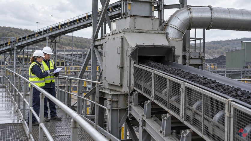
Check dust at the transfer point and beyond
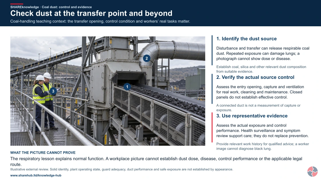
Full written explanation
Coal-handling teaching context: the transfer opening, control condition and workers’ real tasks matter.
- Identify the dust source. Disturbance and transfer can release respirable coal dust. Repeated exposure can damage lungs; a photograph cannot show dose or disease.
- Verify the actual source control. Assess the entry opening, capture and ventilation for real work, cleaning and maintenance. Closed panels do not establish effective control.
- Use representative evidence. Assess the actual exposure and control performance. Health surveillance and symptom review support care; they do not replace prevention.
Change the pathway
- Retain the material uncertainty. Establish coal, silica and other relevant dust composition from suitable evidence.
- No visible cloud is not an all-clear. A connected duct is not a measurement of capture or exposure.
- Keep clinical decisions distinct. Provide relevant work history for qualified advice; a worker image cannot diagnose black lung.
Evidence check: The respiratory lesson explains normal function. A workplace picture cannot establish dust dose, disease, control performance or the applicable legal route.
Full current mine, non-mine, historical and jurisdiction distinctions
Primary references: HSE: mine-law scope and amended dust provisions (GB) · HSE: Mines Regulations guidance, amended2020 (GB) · HSE: task-specific COSHH assessment (GB)
Open the complete HD image (new tab)Download image
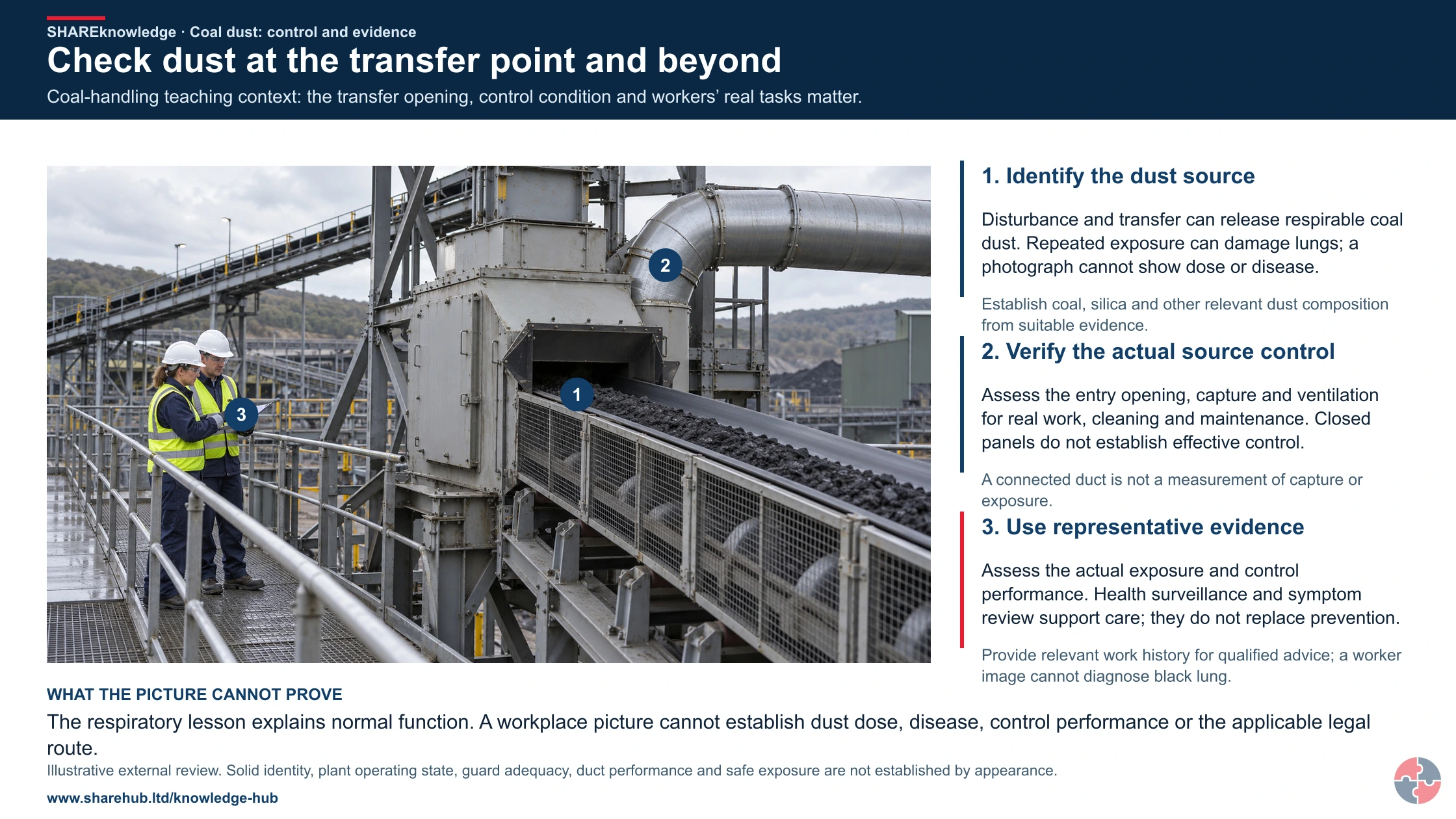
Full current mine, non-mine, historical and jurisdiction distinctions
Primary references: HSE: mine-law scope and amended dust provisions (GB) · HSE: Mines Regulations guidance, amended2020 (GB) · HSE: task-specific COSHH assessment (GB)

Identify the setting
Distinguish current mine, non-mine and historical work. Establish materials, tasks and applicable controls rather than infer a current mine duty from a photograph.
Source and route
Assess enclosure, capture and ventilation for the actual source and people’s positions, including cleaning and maintenance.
Verify and care
Use suitable exposure evidence and act on failed controls. Report symptoms and provide the relevant work history for qualified assessment.
The sketch is not an X-ray, a dust-dose estimate or a diagnosis. No visible dust does not prove adequate control.
What are the key facts about black lung at work?
The key facts are that black lung is caused by respirable coal mine dust, develops slowly, damages lung tissue and is best prevented by controlling dust before it is breathed in.
This page is workplace guidance, not medical, legal or HR advice. Persistent cough, breathlessness, chest tightness, abnormal lung-function results or concern after coal mine dust exposure should be reported through workplace arrangements and assessed through GP, respiratory or occupational health routes.
What signs matter first?
- Persistent cough, breathlessness on exertion, reduced stamina, chest tightness or repeated chest infections.
- Symptoms appearing years after exposure, including after retirement or a move away from mining work.
- Any abnormal health-surveillance, lung-function or chest-imaging result in someone with coal mine dust exposure.
What should happen at work?
- Review coal dust and mixed dust exposure, including respirable dust, silica content, ventilation, water suppression and housekeeping.
- Check exposure monitoring, health surveillance, respiratory protective equipment and maintenance evidence.
- Act on symptoms or health-surveillance trends by reducing exposure and reviewing controls for similar workers.
When is medical assessment needed?
- When respiratory symptoms persist, worsen or interfere with work or normal activity.
- When a worker has a history of coal mine dust exposure and new cough, breathlessness or reduced exercise tolerance.
- When occupational health, health surveillance or a clinician identifies possible work-related lung disease.
It is a pneumoconiosis
Pneumoconiosis is a group of lung diseases caused by inhaling and retaining dust in the lungs.
Coal dust can be mixed dust
Coal mine dust may include coal particles, rock dust, silica-containing dust, diesel particulate and other respiratory irritants depending on the work.
Symptoms can appear late
HSE notes that pneumoconiosis has a long delay between exposure and onset, so current cases may reflect past conditions.
Exposure control comes first
Respiratory protective equipment is not a substitute for controlling dust generation, ventilation, suppression and housekeeping.
Health surveillance is a feedback system
Surveillance should detect early harm and trigger action on workplace controls, not simply produce a medical file.
Records need long life
Dust exposure and health records may matter years later because respiratory disease can have long latency.
What is coal worker’s pneumoconiosis?
Coal worker’s pneumoconiosis is a work-related lung disease caused by inhaling respirable coal mine dust that is retained in the lungs.
The condition is part of the pneumoconiosis family of diseases. In simple terms, very fine dust reaches deep into the lungs, cannot be cleared effectively and can lead to tissue reaction and scarring. The person may have few symptoms early on, but disease can progress and may eventually affect breathing, stamina and work ability.
The term black lung is commonly used because of the association with coal dust. In real workplaces, the exposure picture is often mixed: coal dust may be present alongside stone dust, respirable crystalline silica, diesel engine exhaust, blasting products or other airborne contaminants. A competent assessment therefore looks at the actual dust cloud and work process, not only the job title.
A public-facing workplace page should be clear about the boundary: only a competent clinician can diagnose the disease. The workplace role is to prevent exposure, keep evidence, listen to symptoms and make sure workers are referred through appropriate health routes when concerns arise.
| Element | Explanation | Workplace meaning |
|---|---|---|
| Main cause | Repeated or intense inhalation of respirable coal mine dust. | Assess and control dust at source. |
| Disease type | Pneumoconiosis: dust retained in the lungs causing tissue damage. | Treat as serious occupational lung disease. |
| Latency | Symptoms or diagnosis may occur years after exposure. | Keep long-term exposure and health records. |
| Severity | May be simple or progress to complicated disease such as progressive massive fibrosis. | Act on early indicators and prevent further exposure. |
| Related hazards | Silica, diesel particulate and other mine dust constituents may add risk. | Assess mixed exposures and not coal dust alone. |
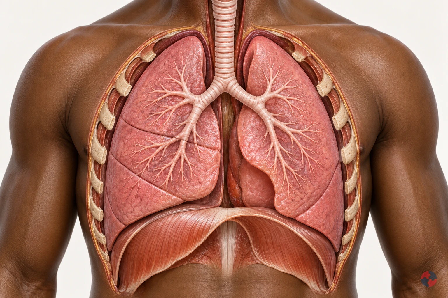

How do the lungs work, and how can workplace exposures affect breathing?
Interactive anatomy · Respiratory tract and lungs · Updated 2026-10-23
Read all anatomy and process explanations without the viewer
Simplified education, not diagnosis, a lung-function or oxygen-saturation test, an exposure calculator or a fitness-to-work assessment. No breath-holding challenge, breathing-rate score or personal health-data collection. Ordinary particle filters do not protect against every gas or oxygen deficiency. COPD and coal-dust topics may reuse normal anatomy only until separate mechanisms are reviewed.
Nose and mouth
Air enters and leaves here. The nose and mouth connect the outside air with the respiratory passages. Air moves through these openings on both the inward and outward breath. Follow the route towards the windpipe before looking at the smaller branches. The illustrated flow is air, a mixture of gases, rather than pure oxygen passing directly into blood. Frimley Health NHS Foundation Trust: How your lungs work · Central and North West London NHS Foundation Trust: How do we breathe?
Trachea
The windpipe carries air into the chest. The trachea, commonly called the windpipe, carries air down towards the chest. It divides into the two main bronchi that supply the lungs. Locate this shared passage before following either branch. Its role is to conduct air towards the gas-exchange regions and provide an outward route for the next exhalation. Frimley Health NHS Foundation Trust: How your lungs work
Bronchi
A main branch carries air into each lung. The two main bronchi take air from the windpipe into the right and left lungs. They divide into progressively smaller branches, distributing the breath through the lungs. Use the split as a landmark before following one highlighted route. These are air passages, distinct from the blood vessels that run through the same organs. Frimley Health NHS Foundation Trust: How your lungs work · University Hospitals of North Midlands NHS Trust: My lung surgery: about your lungs
Bronchioles
Smaller branches distribute air. Bronchioles are smaller branches of the airway system inside the lungs. Their branching routes lead towards clusters of alveoli, where the breath reaches the gas-exchange surface. Follow one branch to its end, using the others as context. This simplified route represents part of a much larger network supplying the lungs together. Frimley Health NHS Foundation Trust: How your lungs work
Alveoli
Tiny air sacs provide a gas-exchange surface. Alveoli are tiny air sacs at the ends of the small airway routes. Their close relationship with surrounding capillaries allows oxygen to pass towards blood and carbon dioxide towards alveolar air. Connect the highlighted cluster with the enlarged gas-exchange view. The enlargement changes the scale so the air space and neighbouring blood vessel can be distinguished. Frimley Health NHS Foundation Trust: How your lungs work · Central and North West London NHS Foundation Trust: How do we breathe?
Air–blood barrier
A thin boundary separates air from blood. The thin alveolar and capillary walls separate air in the sac from blood in its neighbouring vessel. Oxygen and carbon dioxide cross this boundary in opposite directions. Locate the tissue between the two spaces in the enlarged view. Blood cells remain in the vessel; the model does not represent an open connection through which air and blood mix. Central and North West London NHS Foundation Trust: How do we breathe?
Pulmonary capillaries
Small blood vessels pass beside the air sacs. Pulmonary capillaries are small blood vessels beside the alveoli. Blood moving through them receives oxygen and releases carbon dioxide across the thin exchange boundary, then continues through the circulation. Follow the labelled gas routes separately from the blood cells. These cells stay inside the capillary rather than moving into the air sac. Central and North West London NHS Foundation Trust: How do we breathe?
Diaphragm
This muscle helps make room for a breath. The diaphragm is a broad, dome-shaped breathing muscle beneath the lungs. It contracts and flattens downwards during inhalation, helping increase the space in the chest. During a quiet exhalation it relaxes towards its dome shape. Compare it with the rib cage: their changing positions help move air without the lung tissue acting as a contracting muscle. Cambridge University Hospitals NHS Foundation Trust: Breathing techniques to ease breathlessness: muscles involved
Rib cage and breathing muscles
Chest movement changes the space around the lungs. The rib cage protects the chest organs and moves with the breathing muscles. Expansion upwards and outwards helps increase chest space during inhalation; relaxation reduces that space during a quiet exhalation. Compare this movement with the diaphragm below the lungs. Together these structures provide the mechanics that allow air to move through the airways. Central and North West London NHS Foundation Trust: How do we breathe? · Cambridge University Hospitals NHS Foundation Trust: Breathing techniques to ease breathlessness: muscles involved
Pleura
Two thin layers surround the lungs and line the chest. The pleura has two thin layers around the lungs and inside the chest wall. A small amount of fluid between them allows the surfaces to slide with less friction as breathing changes the chest space. Locate this outer covering separately from the internal air sacs; it is not the thin air–blood exchange boundary. Blackpool Teaching Hospitals NHS Foundation Trust: Pleural clinic
Open the complete process transcript
The process, step by step
1 · Make space
Breathing in begins with a change in the space inside the chest. The diaphragm, a dome-shaped muscle beneath the lungs, contracts and moves downwards. Muscles around the ribs help lift the chest upwards and outwards. As the lungs expand with this changing space, air can enter through the airways. Locate the diaphragm and rib cage together rather than treating the lungs as muscles that pull themselves open. The model separates the movement into a teaching step so you can follow its purpose. In the body, chest movement, airflow and blood circulation are connected parts of a continuing process. Central and North West London NHS Foundation Trust: How do we breathe? · Cambridge University Hospitals NHS Foundation Trust: Breathing techniques to ease breathlessness: muscles involved
What to watch: Locate the diaphragm beneath the lungs and the rib cage around them; compare their positions with the space occupied by the lungs.
Why it matters: Changing the chest space draws air into the lung passages. The breathing muscles supply the movement, while the lungs expand with the chest to receive the next breath.
Explore this step: Select the diaphragm and rib cage in turn, then explain how their movement helps air enter without the lungs contracting like muscles.
2 · Move air
Air entering through the nose or mouth travels down the trachea, or windpipe, towards the chest. The trachea divides into the two main bronchi, with one route supplying each lung. Follow this continuous passage in the whole-chest view before moving into the smaller branches. The moving air is a mixture of gases, including oxygen; it is not a stream of pure oxygen travelling straight into the blood. These larger airways carry the breath towards the places where gas exchange happens. The same connected passages also provide the outward route when air is breathed out. Frimley Health NHS Foundation Trust: How your lungs work
What to watch: Start at the nose and mouth, follow the trachea downwards and find the split into the right and left main bronchi.
Why it matters: The larger passages distribute air to both lungs and connect the outside air with the smaller branches inside. Gas exchange takes place further along the route, rather than within the windpipe.
Explore this step: Trace the route to each lung and distinguish the tubes carrying air from the separate vessels carrying blood.
3 · Reach the air sacs
Inside each lung, the bronchi divide into smaller branches called bronchioles. These passages distribute air towards clusters of tiny air sacs, the alveoli. Follow one branch in the close-up and connect its end with an air-sac cluster, using the other branches as surrounding context. These routes distribute the breath throughout the lungs rather than supplying only one working air sac. A real lung contains many branching routes and air sacs operating together. This distribution brings air close to a large network of small blood vessels, preparing for the exchange shown in the next view. Frimley Health NHS Foundation Trust: How your lungs work
What to watch: Follow one smaller airway to the cluster of air sacs at its end, keeping the other branches as surrounding context.
Why it matters: Branching delivers air throughout the lungs instead of to one large chamber. The many air sacs bring the inhaled gases close to blood vessels where oxygen and carbon dioxide can cross.
Explore this step: Trace one complete branch to its air sacs, then explain how the many branches distribute air throughout the lungs.
4 · Change scale
The lesson now changes from a branching airway to an enlarged air–blood interface. An alveolus contains air; beside it runs a capillary containing blood. Their very thin walls form the boundary across which gases can move. Locate the air space, the separating tissue and the inside of the vessel before following any gas symbols. The magnified view represents a tiny part of the same lung, not a separate organ or a hole that joins air and blood into one space. The enlargement helps explain how the two systems can exchange gases while keeping their contents separate. Central and North West London NHS Foundation Trust: How do we breathe?
What to watch: Find the air inside the enlarged alveolus, the thin separating boundary and the neighbouring capillary with blood cells inside it.
Why it matters: A close boundary allows gases to cross between alveolar air and capillary blood. Keeping the two spaces separate lets ventilation and circulation cooperate without blood cells entering the air sac.
Explore this step: Identify the air space and blood-vessel space, then point to the boundary each gas must cross between them.
5 · Exchange gases
At the air–blood boundary, oxygen moves from the alveolar air into the capillary blood. At the same time, carbon dioxide carried back from the body moves from the blood into the air sac. The two gases cross in opposite directions; blood cells stay within the vessel. Follow each labelled symbol separately so that exchanging gases is not confused with mixing the air and blood together. Blood then carries oxygen onwards towards the heart and the body’s tissues. The teaching symbols simplify continuous gas movement, and their size, colour and speed do not represent a measurement. Central and North West London NHS Foundation Trust: How do we breathe?
What to watch: Follow the O₂ symbol towards the capillary and the CO₂ symbol towards the air sac, while locating the blood cells within the vessel.
Why it matters: Oxygen can be carried onwards for use by the body, while carbon dioxide returns to the airways for removal. Both transfers connect the breath with the continuing circulation of blood.
Explore this step: Trace the two gas routes in opposite directions and explain why a blood cell should remain inside the capillary.
6 · Breathe out
During a quiet breath out, the diaphragm and other breathing muscles relax. The diaphragm returns towards its dome shape, chest space decreases and air moves back along the airways to the nose or mouth. This outgoing air carries carbon dioxide that crossed from the blood into the alveoli. Trace the route in reverse without reversing the direction of blood circulation: oxygen-carrying blood continues towards the heart while another breath prepares to enter. The model presents a single cycle to explain the relationship. It does not show breathing as a series of separate events in which circulation stops between steps. Central and North West London NHS Foundation Trust: How do we breathe? · Cambridge University Hospitals NHS Foundation Trust: Breathing techniques to ease breathlessness: muscles involved
What to watch: Compare the diaphragm and chest with the inhalation step, then follow the outward airway route from the lungs to the nose or mouth.
Why it matters: Exhalation removes air containing carbon dioxide from gas exchange. Meanwhile, blood circulation continues carrying oxygen onwards, linking this breathing cycle with the needs of the rest of the body.
Explore this step: Trace the outward air route and the onward blood route separately, then describe why they are connected processes with different pathways.
Read the practice situations and all answer explanations
Sources and how the model is made
The opening images are AI-generated anatomical illustrations. The interactive 3D models and labelled SVG diagrams use original procedural artwork. These teaching images are not medical scans or diagnostic tools. Source references support the learner explanations and do not grant artwork rights.
- Frimley Health NHS Foundation Trust: How your lungs work — Normal airway branching, air sacs and capillary gas exchange; factual text only.
- Central and North West London NHS Foundation Trust: How do we breathe? — Thin alveolar/capillary walls, opposing gas transfer, circulation and chest mechanics. No illustrations or exercises copied.
- Cambridge University Hospitals NHS Foundation Trust: Breathing techniques to ease breathlessness: muscles involved — Diaphragm movement and breathing-muscle context only; this module does not prescribe breathing exercises.
- HSE: Health risks from welding — Welding-related airway irritation, possible deeper injury and irritant asthma; not a universal pattern for every gas.
- HSE: Asthma: an overview — Sensitisation, later triggering and work aggravation. Legacy labelling language and statistics are not reused.
- HSE: Advice for employers: occupational asthma — Exposure prevention, controls and health surveillance only; legacy legislation wording is not reused.
- NHS: Silicosis — Silica-associated inflammation/fibrosis, varying latency, irreversible damage and possible progression after exposure stops.
- HSE: Why is asbestos dangerous? — Asbestosis, mesothelioma and pleural disease are distinct; long disease latency.
- HSE: Construction dust: cutting paving blocks, kerbs and flags — Task design, source suppression/extraction, suitable RPE and checking controls; not a generic dust safe-dose rule.
- NHS: Shortness of breath — Emergency recognition of severe breathing difficulty and 999; emergency advice is outside the activity.
- NHS: Asthma — Narrowed/swollen airways, symptoms and clinical diagnosis; no medication doses or treatment plan reproduced.
- University Hospitals of North Midlands NHS Trust: My lung surgery: about your lungs — Right/left lung lobe counts and airway anatomy; no source image copied.
- Blackpool Teaching Hospitals NHS Foundation Trust: Pleural clinic — Pleural layers and the small lubricating fluid layer outside lung tissue; no procedural teaching included.
- HSE: Asphyxiation hazards in welding and allied processes — Oxygen-deficient spaces and need for independent air supply; no unprotected entry or rescue.
- HSE: Respiratory protective equipment at work — RPE selection must match the contaminant and conditions; filtering protection is not a supply of oxygen.
- HSE: Flour dust and risks to bakers — Flour sensitisation and practical reduction of dusty handling; exposure-limit numbers are not reused.
- Health and Safety Executive: Asbestos essentials — Task planning, training and competent asbestos work arrangements for the maintenance exposure example; not permission to disturb unknown material or a licence/classification decision.
- Health and Safety Executive: Welding fume: protect your workers — Alternative joining, fume reduction, LEV, suitable RPE and maintained controls for the welding-fume example. No unprotected exposure or rescue procedure.
What is different about coal mine dust?
Coal mine work can generate respirable coal dust and a separate respirable crystalline silica component when rock is cut; assess both the fraction and task.
Extraction, cutting, roof work, crushing, conveying and cleanup can create very different concentrations. Inhalable dust and respirable dust are distinct measurement fractions, and invisible respirable particles can remain hazardous when the air looks clear. Coal worker’s pneumoconiosis is associated with coal dust, while silica may cause silicosis and contribute to mixed-dust disease. Do not classify a coal-handling site automatically as a below-ground mine: the specific regulations depend on the operation.
On a small screen, scroll this table sideways to read all columns.
| Task | Likely concern | Control check |
|---|---|---|
| Cutting coal | Coal mine dust | Suppression, ventilation and breathing-zone sampling |
| Cutting rock or roof | Respirable silica as well as coal dust | Separate silica assessment and sampling |
| Conveying or cleanup | Resuspended inhalable and respirable dust | Enclosure, extraction and safe cleaning |
How does coal dust damage the lungs?
Respirable coal mine dust retained in the lungs can produce tissue changes; more extensive disease can cause scarring and loss of lung function.
Healthy lungs have defence systems that trap and clear many particles. Respirable coal mine dust can bypass the upper airway and reach deeper lung tissue. Accumulated coal mine dust can produce macules and nodules; more extensive disease may develop into progressive massive fibrosis.
Simple pneumoconiosis may cause few symptoms at first, which makes monitoring and health surveillance important. Complicated disease can involve larger areas of scarring, reduced gas exchange, breathlessness and serious disability. Smoking is not the cause of coal worker’s pneumoconiosis, but it can worsen respiratory symptoms and overall lung health, so it should be considered in health advice without distracting from workplace exposure control.
| Stage | What happens | Why control matters |
|---|---|---|
| Inhalation | Fine respirable dust enters deep lung regions. | Control dust before it reaches the breathing zone. |
| Retention | Particles remain in lung tissue and are not cleared effectively. | Repeated exposure increases body burden. |
| Tissue reaction | Macules and nodules can form around retained coal mine dust. | Early symptoms may be absent. |
| Fibrosis | Scarring can stiffen lung tissue and reduce function. | Damage may be long-term and disabling. |
| Complication | Progressive massive fibrosis or other respiratory disease may develop. | Health surveillance and exposure reduction are critical. |
The IIAC2023 pneumoconiosis review describes early coal macules around terminal bronchioles, which may coalesce into the larger abnormalities of progressive massive fibrosis. Diagnosis combines exposure history, imaging and exclusion of similar conditions. Simple CWP generally causes no symptoms or disability; a diagnosis does not by itself establish functional impairment. The report’s benefit recommendations are advisory: use the current scheme guidance for an actual claim.
What symptoms can suggest coal worker’s pneumoconiosis?
Simple CWP is generally asymptomatic. More extensive respiratory disease can affect breathing and exercise tolerance; cough or chest tightness also need assessment for other causes.
The absence of symptoms does not prove that exposure is controlled. Coal worker’s pneumoconiosis can be detected after long latency, and some workers may first notice difficulty with stairs, long walks, heavy work or respirator use rather than obvious illness.
Workers should be encouraged to report changes early and to describe their occupational history clearly to clinicians. Managers should not dismiss respiratory symptoms as age, fitness or smoking without considering work exposure history and health-surveillance findings.
| Sign or symptom | What it may indicate | Workplace response |
|---|---|---|
| Persistent cough | Possible chronic airway irritation or occupational lung disease. | Record concern and refer through OH/GP route. |
| Breathlessness on exertion | Reduced lung function or respiratory disease. | Review exposure and fitness for dusty/physical work. |
| Reduced stamina | Functional impact from respiratory impairment or other disease. | Consider work demands and health advice. |
| Chest tightness | Possible respiratory or other medical issue. | Encourage medical assessment route. |
| Abnormal surveillance result | Possible early health change or control failure. | Act on occupational health advice and review controls. |
Cough, tightness or reduced stamina are nonspecific and may reflect another or coexisting condition; they do not diagnose CWP. New or worsening concerns require clinical assessment with the actual exposure history. Symptom-free workers still need effective dust prevention and the applicable surveillance arrangements.
Which work activities create higher risk?
Higher-risk activities are those that generate or disturb respirable coal mine dust or mixed dust in poorly controlled work areas.
In Great Britain, active coal mining is far smaller than it once was, but the lesson remains relevant to mining, tunnelling, mineral extraction, legacy plant, coal handling and any work disturbing coal-containing deposits. Historical exposure also matters because claims and diagnoses may relate to work done many years earlier.
Risk is shaped by the dustiness of the task, particle size, silica content, ventilation, duration, frequency, enclosure, maintenance standard, cleanliness, worker position and whether controls are used consistently. A short but extremely dusty task can be important, and a low-level task can still matter if it is repeated for years.
| Work situation | Why risk can increase | What to check |
|---|---|---|
| Underground coal extraction | Enclosed work, mechanical cutting and variable ventilation can create respirable dust. | Ventilation, sprays, monitoring and operator position. |
| Coal crushing and screening | Material impact and transfer can release fine dust. | Enclosure, extraction, suppression and maintenance. |
| Conveyor maintenance | Settled dust is disturbed around belts, transfer points and structures. | Isolation, cleaning method and RPE programme. |
| Legacy coal plant or storage | Old deposits can be disturbed during refurbishment or demolition. | Survey, cleaning plan, monitoring and contractor control. |
| Mixed mineral strata work | Coal work can also disturb silica-containing rock. | RCS assessment, monitoring and health surveillance. |
How should mine dust be measured and acted on?
Use a competent sampling plan for representative tasks. Regulation 45’s sampling and action-level loop is for respirable dust; assess silica separately under COSHH.
GB Mines Regulations 2014 regulation 45 applies below ground in coal mines. It requires recorded assessment of inhalable dust risk, prevention or reduction so far as reasonably practicable, and suitable sampling of respirable dust. If the respirable-dust action level is exceeded, the operator must determine why, take remedial action and sample again to check effectiveness. The 2020 amendment removed silica references from regulation 45’s sampling/action-level paragraphs and revoked the below-ground COSHH exception. Respirable crystalline silica is assessed, monitored where appropriate and controlled under COSHH and its current workplace exposure limit. Verify current units, averaging periods and legal thresholds from the live instrument and EH40 with a competent hygienist; this page does not supply a universal coal-dust limit.
On a small screen, scroll this table sideways to read all columns.
| Step | Question | Evidence |
|---|---|---|
| Plan | Which worker and worst credible task? | Sampling strategy |
| Measure | Were the fractions and methods appropriate? | Calibrated result and task log |
| Respond | Why did an action level or control fail? | Investigation and action |
| Verify | Did the correction reduce exposure? | Repeat sampling and worker feedback |
How can the hierarchy of controls reduce coal-dust exposure and black-lung risk?
Treat dust generation and inhalation as the harm chain: redesign the cut or transfer, then engineer dust capture, maintain the system and use RPE only for residual exposure.
The HSE five-level hierarchy asks whether elimination, substitution, engineering controls, administrative controls and PPE can reduce risk in that order. ERICPD cross-maps elimination, reduction including substitution, isolation, engineering control, PPE and discipline. These are decision aids, not identical legal ladders. Mining cannot eliminate all coal cutting while still extracting coal; it can remove unnecessary dry cleanup and redesign transfer points. A mine engineer should test whether a less dusty cutting method or pre-wetting is compatible with ground conditions and equipment before work, rather than promising substitution without trial.
At a cutting face, the operator specifies working water sprays and ventilation to move dust away from breathing zones. At a conveyor transfer, an enclosed chute and extraction collector can keep dust inside the system; a supervisor verifies seals, airflow and filter condition. A closed cab or remote control isolates a worker only if filtration, pressure and doors are maintained. Procedures for inspection, cleaning and restricted access sustain these engineered barriers but do not replace them. Stop or restrict a dusty task when sprays, ventilation, enclosure or RPE fail, assess exposure, repair and verify effectiveness before restart.
On a small screen, scroll this table sideways to read all columns.
How can the ERICPD hierarchy of controls reduce coal-dust exposure and black-lung risk?

| Five-level / ERICPD | Specific intervention and feasibility | Owner, check, failure and stop |
|---|---|---|
| Elimination / Eliminate | Remove dry sweeping and compressed-air blowdown from belt maintenance; vacuum or wet-clean before opening the guard. Coal extraction itself cannot be removed without changing the operation. | Maintenance lead verifies method and captured waste; stop cleanup if dry dust clouds form. |
| Substitution / Reduce | Trial a lower-dust cutting or pre-wetted transfer method where geology and machine instructions permit; avoid replacing a coal hazard with uncontrolled silica or water/electrical risk. | Mine engineer approves trial; compare personal dust samples and equipment performance; reject if new risk or no benefit. |
| Engineering / Isolate and Control | Fit enclosed transfer chute, source extraction and maintained filtration; use correctly directed ventilation and water sprays at the face, and sealed filtered cab where suitable. | Operator commissions airflow/spray pattern and monitors worker exposure; stop or restrict work for failed spray, low airflow, leaking chute or cab seal. |
| Administrative / Discipline | Plan sampling, inspection, maintenance, access control, defect escalation and clean/dirty change arrangements; train workers on the actual stop rule. | Supervisor signs pre-use checks and reviews exceedances; hold restart until repair and resampling verify control. |
| PPE / PPE | Specify suitable RPE for residual exposure from the actual dust assessment, fit-test tight-fitting models, check seal, filter and compatibility, and replace/maintain as directed. | Competent assessor records selection; user checks fit before entry; stop if fit, filter or supply fails. |
How can the ISO 45001 hierarchy of controls reduce coal-dust exposure and black-lung risk?

| Five-level / ERICPD | Specific intervention and feasibility | Owner, check, failure and stop |
|---|---|---|
| Elimination / Eliminate | Remove dry sweeping and compressed-air blowdown from belt maintenance; vacuum or wet-clean before opening the guard. Coal extraction itself cannot be removed without changing the operation. | Maintenance lead verifies method and captured waste; stop cleanup if dry dust clouds form. |
| Substitution / Reduce | Trial a lower-dust cutting or pre-wetted transfer method where geology and machine instructions permit; avoid replacing a coal hazard with uncontrolled silica or water/electrical risk. | Mine engineer approves trial; compare personal dust samples and equipment performance; reject if new risk or no benefit. |
| Engineering / Isolate and Control | Fit enclosed transfer chute, source extraction and maintained filtration; use correctly directed ventilation and water sprays at the face, and sealed filtered cab where suitable. | Operator commissions airflow/spray pattern and monitors worker exposure; stop or restrict work for failed spray, low airflow, leaking chute or cab seal. |
| Administrative / Discipline | Plan sampling, inspection, maintenance, access control, defect escalation and clean/dirty change arrangements; train workers on the actual stop rule. | Supervisor signs pre-use checks and reviews exceedances; hold restart until repair and resampling verify control. |
| PPE / PPE | Specify suitable RPE for residual exposure from the actual dust assessment, fit-test tight-fitting models, check seal, filter and compatibility, and replace/maintain as directed. | Competent assessor records selection; user checks fit before entry; stop if fit, filter or supply fails. |
When is health surveillance needed?
Below-ground GB coal mine workers who are or may be exposed to inhalable dust require suitable surveillance unless exposure is not significant; other settings need their own risk-based decision.
Regulation 45(7) places that specific duty on the GB mine operator. Occupational health should determine suitable respiratory surveillance and referral from the risk profile. Surveillance does not replace air sampling or dust control, and a normal check cannot prove a dusty task is safe. If symptoms or abnormal findings arise, protect the worker, review the controls and look for similarly exposed people. An individual clinical record should remain confidential; managers need only necessary fitness and control advice.
On a small screen, scroll this table sideways to read all columns.
| Trigger | Owner | Outcome |
|---|---|---|
| Exposure likely significant | Mine operator and occupational health | Programme and baseline decision |
| Finding or symptom | Occupational health | Clinical referral and work advice |
| Control review | Mine operator | Exposure reduction and verification |
| Record | Health professional and employer | Separate clinical and exposure records |
What records should be kept for coal dust and black lung prevention?
Records should prove exposure was assessed, controls were maintained, workers were trained, health surveillance was provided and findings led to action.
Because pneumoconiosis can appear years after exposure, records need to be durable and retrievable. A business should be able to reconstruct who did what work, where, with which controls, what monitoring showed and what health-surveillance arrangements applied.
COSHH health records are not the same as confidential medical records. Employers need suitable health records and action evidence, while clinical details should remain under appropriate medical confidentiality. This distinction prevents both under-recording and inappropriate sharing.
| Record | Why it matters | Quality standard |
|---|---|---|
| COSHH assessment | Shows dust routes, exposed groups and control decisions. | Specific to task, dust type and controls. |
| Exposure monitoring | Shows airborne dust levels and whether controls are adequate. | Representative, interpreted and actioned. |
| Ventilation and suppression checks | Shows engineering controls were working. | Routine checks, defects and repairs recorded. |
| RPE records | Shows respirator suitability, fit, maintenance and issue. | Face-fit, APF selection, cleaning and replacement. |
| Health surveillance health records | Shows inclusion, date, outcome category and review needs. | Long-term retention and confidential handling. |
| Training and competence | Shows workers and supervisors understood controls. | Role-specific, refreshed and checked in practice. |
Which law and support routes apply?
GB and NI have separate mining instruments. GB regulation 45 samples respirable dust; silica falls under COSHH. Benefit and compensation routes differ from incident reporting.
For a below-ground GB coal mine, Mines Regulations 2014 regulation 45 sets inhalable-dust assessment/control, respirable-dust sampling and health-surveillance duties; silica is separately assessed under COSHH. HSE confirms older coal-dust ACOP L145 is withdrawn. In NI, the Mines Regulations (NI) 2016 and 2022 amendment interact with NI COSHH, notably for silica below ground. Check the actual NI instrument with a mine specialist. A former miner may be eligible for GB Industrial Injuries Disablement Benefit prescribed disease D1 or the current Coal Industry Pneumoconiosis Compensation Scheme if criteria are met; accepting a scheme payment prevents a court claim, so obtain legal advice first. NI benefit routes differ. Do not present IIDB eligibility, a compensation scheme or a RIDDOR report as automatic.
On a small screen, scroll this table sideways to read all columns.
| Question | Great Britain | Northern Ireland |
|---|---|---|
| Mine dust | MR14 regulation 45 below ground; COSHH where applicable | Mines (NI) 2016 as amended; NI COSHH |
| Pneumoconiosis reporting | Not separately on current GB RIDDOR diagnosed-disease list | Check NI reporting rules against diagnosis and work facts |
| Benefits | IIDB D1 subject to occupation and diagnosis criteria | NI IIDB process and criteria |
| Coal compensation | Current GOV.UK scheme criteria and election against court claim | Seek local legal/benefit advice; do not import GB scheme terms |
How should black lung risk appear in the risk register?
Black lung risk should appear as a respiratory disease scenario linked to coal dust generation, exposed groups, controls, monitoring, surveillance and long-term records.
The risk register should describe the exposure route, not only the disease label. For example: respirable dust from conveyor cleaning, mixed dust from cutting in coal measures, coal handling in enclosed plant, or maintenance inside dusty equipment.
Each entry should identify the barrier that prevents exposure, the assurance evidence and the trigger for review. A monitoring result above expectations, a failed ventilation check, a health-surveillance trend or a worker report of symptoms should lead to action.
| Scenario | Risk route | Controls | Assurance |
|---|---|---|---|
| Conveyor transfer dust | Fine dust released at transfer points and breathed by operators. | Enclosure, extraction, suppression and cleaning. | Air monitoring and maintenance records. |
| Maintenance in dusty plant | Settled dust disturbed during access and repair. | Isolation, pre-cleaning, wet methods, RPE and permit. | Permit, evidence log and supervisor check. |
| Vehicle movement | Settled dust re-suspended along routes. | Roadway control, water suppression and cab filtration. | Dust inspections and cab filter maintenance. |
| Mixed coal and silica dust | Coal work disturbs silica-containing strata or rock dust. | RCS assessment, monitoring and health surveillance. | Occupational hygiene report and COSHH review. |
| Historical exposure | Former workers develop symptoms years later. | Record retention, exposure history and OH referral route. | Archived monitoring and health records. |
How could Crownway Fuels Ltd manage coal dust exposure risk?

Crownway Fuels Ltd would manage coal dust exposure by treating it as a respiratory health risk requiring competent assessment, engineered control and long-term evidence.
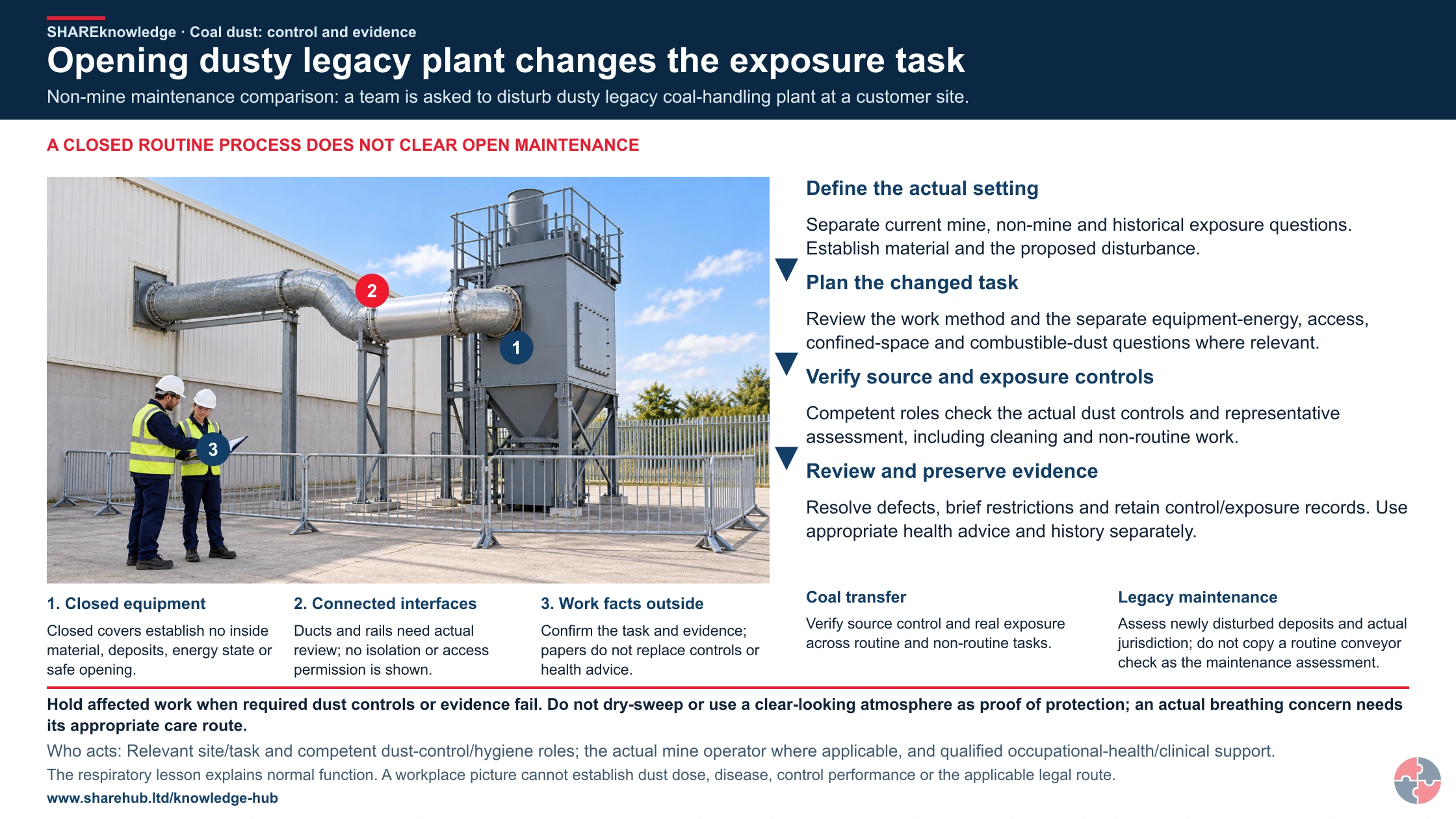
Opening dusty legacy plant changes the exposure task
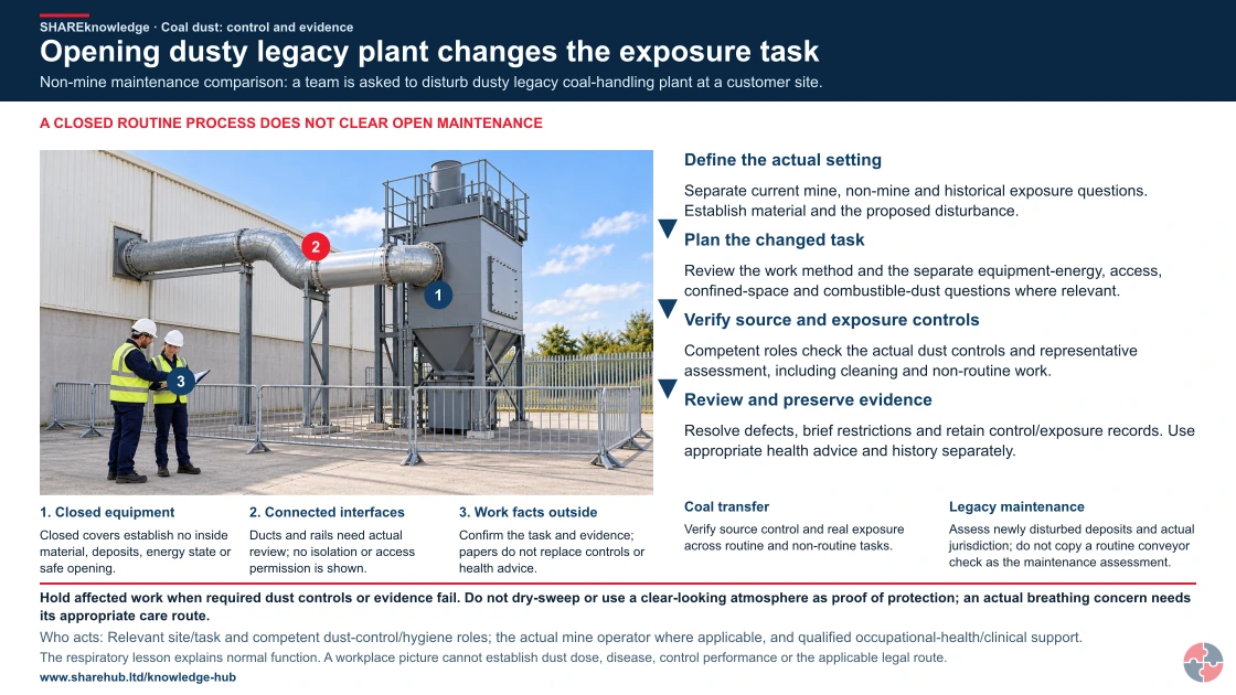
Full written explanation
An illustrative external equipment review shows a whole closed collector and hopper, connected duct, freestanding work boundary and normal colleagues comparing inert facts outside. Picture key: 1 identifies closed equipment; 2 identifies connected interfaces; 3 identifies work facts outside. The scene is generic physical context before a proposed maintenance task. It is not identified as an actual coal plant, mine, legacy installation or customer site. Contents, deposits, material composition, dust fractions, plant age, operating and energy state remain unknown. Closed panels, ducts and rails do not demonstrate isolation, safe access, capture, combustible-dust protection or permission to open. The complete four-check review retains the actual setting, changed work, source/exposure-control evidence and separately preserved work/health records. Independent headings are unnumbered in one ordered review band. A closed routine process does not clear open maintenance; the full hold, owners, evidence limits and both workplace cases remain. Routine transfer checks cannot stand in for the proposed disturbance assessment. The existing normal-only respiratory lesson remains distinct from any diagnosis, dust dose, surveillance result or historical attribution. Urgent breathing concerns and appropriate care do not wait for the task review or records. No panel opening, entry, disturbance, cleaning, dry sweeping, dust cloud, exposure, sampling, anatomy, harm, symptoms, clinical or emergency demonstration is shown. Use the complete source for its current mine, non-mine, historical, GB/NI, silica, clinical, benefit/reporting and confidentiality qualifications.
Non-mine maintenance comparison: a team is asked to disturb dusty legacy coal-handling plant at a customer site.
- Define the actual setting. Separate current mine, non-mine and historical exposure questions. Establish material and the proposed disturbance.
- Plan the changed task. Review the work method and the separate equipment-energy, access, confined-space and combustible-dust questions where relevant.
- Verify source and exposure controls. Competent roles check the actual dust controls and representative assessment, including cleaning and non-routine work.
- Review and preserve evidence. Resolve defects, brief restrictions and retain control/exposure records. Use appropriate health advice and history separately.
When the evidence does not fit: Hold affected work when required dust controls or evidence fail. Do not dry-sweep or use a clear-looking atmosphere as proof of protection; an actual breathing concern needs its appropriate care route.
Who acts: Relevant site/task and competent dust-control/hygiene roles; the actual mine operator where applicable, and qualified occupational-health/clinical support.
Changed workplace
Coal transfer: Verify source control and real exposure across routine and non-routine tasks.
Legacy maintenance: Assess newly disturbed deposits and actual jurisdiction; do not copy a routine conveyor check as the maintenance assessment.
Evidence check: The respiratory lesson explains normal function. A workplace picture cannot establish dust dose, disease, control performance or the applicable legal route.
Full current mine, non-mine, historical and jurisdiction distinctions
Primary references: HSE: mine-law scope and amended dust provisions (GB) · HSE: Mines Regulations guidance, amended2020 (GB) · HSE: task-specific COSHH assessment (GB)
Open the complete HD image (new tab)Download image
Full current mine, non-mine, historical and jurisdiction distinctions
Primary references: HSE: mine-law scope and amended dust provisions (GB) · HSE: Mines Regulations guidance, amended2020 (GB) · HSE: task-specific COSHH assessment (GB)
Crownway Fuels Ltd
A fuel distribution and industrial maintenance business that occasionally handles legacy solid-fuel systems, dusty plant, contractors and enclosed transfer equipment.
Crownway is not a coal mine operator, so it would not attempt to manage mine-specific risks without specialist support. Its relevant scenario is legacy coal or coke handling equipment at a customer site, where contractors are asked to clean, isolate, remove or refurbish dusty conveyors, hoppers or silos. Before work starts, Crownway commissions a competent assessment of dust type, silica content, confined-space potential, explosive-dust potential and respiratory health risk.
The risk register separates immediate project controls from long-term occupational health controls. Immediate controls include pre-cleaning by controlled methods, wet suppression where suitable, segregation, permits, isolation, negative-pressure or extraction arrangements, Type H vacuum cleaning, suitable RPE and welfare. Long-term controls include training, exposure monitoring, health-surveillance decisions, contractor competence, document retention and management review.
Crownway checks the work in three layers. Frontline supervisors use binary checks for suppression, extraction, RPE fit evidence, housekeeping and exclusion zones. HSE specialists sample the permit pack, COSHH assessment, monitoring plan and contractor competence. A periodic assurance review checks whether dusty legacy work is being captured in procurement, project planning, legal register and incident learning rather than treated as one-off cleaning.
| Management area | How Crownway applies it | Evidence and improvement |
|---|---|---|
| Assess and analyse | Identifies coal dust, silica, confined-space, combustible-dust and respiratory-health routes before work starts. | COSHH assessment, occupational-hygiene brief and project risk register. |
| Implement controls | Uses enclosure, wet suppression, extraction, no dry sweeping, RPE, isolation and permit controls. | Permit pack, control checks, photographs and equipment inspection records. |
| Train and supervise | Briefs workers and contractors on dust hazards, RPE, symptoms, hygiene and stop-work triggers. | Training records, toolbox talk, face-fit certificates and supervisor observations. |
| Monitor and check | Uses exposure monitoring where needed and samples control performance during work. | Air monitoring report, maintenance logs and action close-out. |
| Health surveillance | Uses occupational health advice to decide who needs surveillance and how symptoms are referred. | Surveillance register, health records and anonymised trend review. |
| Learn and improve | Feeds findings into contractor standards, procurement, method selection and future project planning. | Lessons learned, revised templates and management review minutes. |
What should each role learn and demonstrate?
Learning must match the decisions people make, from recognising danger to checking that controls work.
Briefings should tell people how to act, but attendance alone does not show competence. Assess the real decision or task, record who assessed it and revisit after a material change, failed control or incident. A local authorisation is task-specific; no universal external certificate is implied.
On a small screen, scroll this table sideways to read all columns.
| Role and learning | Demonstration and assessor | Record and reassessment trigger |
|---|---|---|
| Mine operator: dust fractions and duties. | Identify regulation 45 inhalable-dust assessment, respirable-dust sampling, separate COSHH silica assessment and surveillance triggers. | Reviewed site plan and sampling programme. |
| Supervisor: controls in use. | Inspect sprays, ventilation, enclosure and cleaning during a real dusty task. | Defect and restart record. |
| Occupational health: findings. | Explain referral, work protection and confidential feedback. | Programme protocol and action log. |
| Claims adviser: eligibility boundary. | Explain that IIDB D1 and coal compensation require criteria and legal advice before scheme acceptance. | Checked signposting sheet. |
How does this topic support NEBOSH study and professional development?
This topic supports study by linking hazardous substances, occupational lung disease, long latency, exposure monitoring, health surveillance and management evidence.
For general certificate learners, the core is understanding that dusts can be hazardous substances under COSHH, that inhalation is a key route into the body and that control should follow the hierarchy before relying on PPE. Learners should also be able to explain why health surveillance may be needed and why absence of symptoms does not prove absence of harm.
For diploma or professional development, the topic is more analytical: assess mixed dust, select monitoring methods, interpret trends, challenge weak controls, handle long-latency records, consult occupational health and explain how findings should change procurement, maintenance, supervision and management review.
| Learning area | What to know | Scenario application |
|---|---|---|
| Disease mechanism | Respirable dust reaches deep lungs and can cause fibrosis. | Explain why visible housekeeping alone is not enough. |
| COSHH assessment | Dusts and WELs must be assessed and controlled. | Build an assessment for a dusty plant task. |
| Monitoring | Air monitoring checks exposure and control effectiveness. | Decide when specialist sampling is needed. |
| Health surveillance | Surveillance detects work-related health effects and informs control review. | Explain what happens after abnormal trends. |
| Records | Long-latency disease needs durable exposure and health records. | Identify records needed years later. |
How can a management system sustain effective decisions?
Give each hazard or support route an owner, a control, a check and a response to failure.
Audit the mine operator’s dust assessment, source controls, sampling response and surveillance together; keep compensation advice separate from hazard control. An owner should sample actual work and corrective actions, not accept a policy or certificate as evidence that the controls work.
The free GB regulator guide HSE Managing for health and safety (HSG65) explains a plan–do–check–act approach: assign controls and owners, involve workers, check work as done, and correct failures. For Northern Ireland, use HSENI risk assessment guidance for the local duty. A certificate alone does not demonstrate that a control works.
What warning signs show coal dust controls are failing?
Warning signs include visible dust, poor ventilation, dry cleaning, weak RPE management, missing monitoring, symptoms and health-surveillance trends.
A failing system often shows itself before disease is diagnosed. Workers see dust clouds, filters block, sprays are not aligned, cabs are dirty, dust settles in welfare areas, masks are worn over stubble, or supervisors accept dusty shortcuts because the job is behind schedule. Each sign indicates potential harm and poor evidence.
The response should be practical and fast. Stop the uncontrolled exposure route where needed, fix engineering controls, clean using safe methods, repeat monitoring, consult workers, review health surveillance and check similar tasks elsewhere.
| Warning sign | How harm can develop | What to do |
|---|---|---|
| Visible dust cloud | Respirable dust may be reaching the breathing zone. | Stop, review suppression or extraction and monitor exposure. |
| Dry sweeping or compressed air | Settled dust becomes airborne and inhalable. | Use Type H/vacuum or wet methods. |
| Poor ventilation or failed sprays | Dust is not diluted, captured or suppressed. | Repair, test and verify before continuing. |
| RPE not face-fit tested | The mask may leak and give false protection. | Face-fit, train, supervise and maintain. |
| Symptoms normalised | Early disease signs may be missed. | Report, refer and review exposure. |
| Monitoring results not actioned | Known exposure weakness persists. | Assign actions, verify controls and repeat monitoring. |
Chemical hazard symbols: what do they mean?
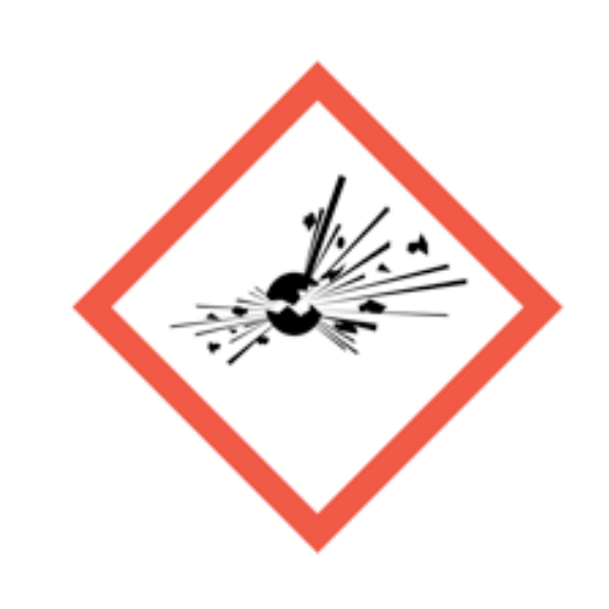
Exploding bomb
Explosive, self-reactive or organic peroxide hazards.
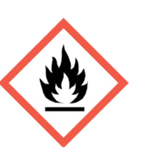
Flame
Flammable, self-heating or pyrophoric substances.
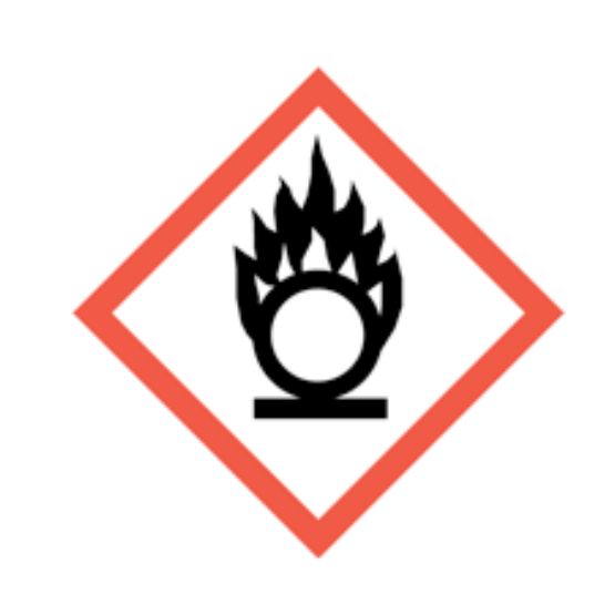
Flame over circle
Oxidising substances that can intensify fire.

Gas cylinder
Gas under pressure.
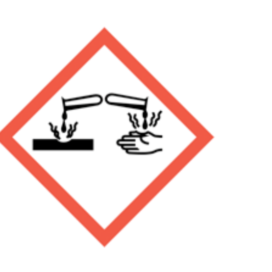
Corrosion
Skin burns, eye damage or corrosive to metals.
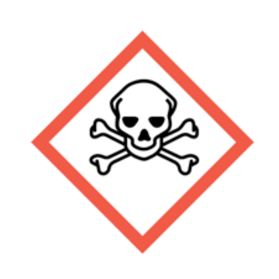
Skull and crossbones
Acute toxicity.
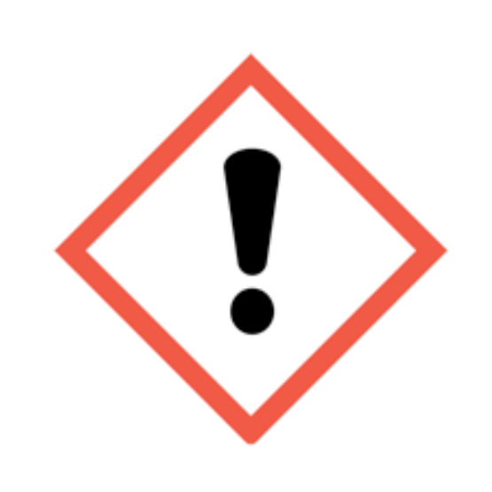
Exclamation mark
Irritation, harmful effects or skin sensitisation; some classifications cover drowsiness, dizziness or ozone-layer hazard. Read the actual hazard statements. Skin sensitisation differs from respiratory sensitisation.
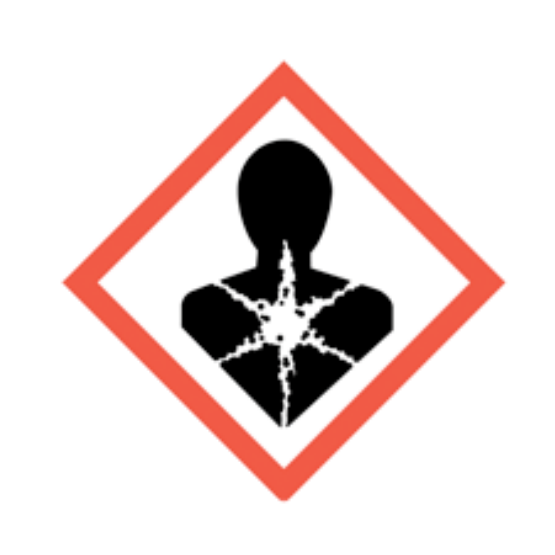
Health hazard
Serious health hazards, including organ damage, aspiration hazard, respiratory sensitisation, cancer and reproductive effects; some can follow a single exposure.
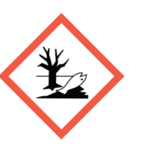
Environment
Hazardous to the aquatic environment.
What acronyms are used on this black lung page?
Acronyms should be explained so respiratory-health and legal-control language remains accessible.
| Acronym | Meaning | Why it matters |
|---|---|---|
| CWP | Coal worker’s pneumoconiosis | The formal disease name often called black lung. |
| COSHH | Control of Substances Hazardous to Health | Main UK regulations for controlling hazardous dust exposure. |
| EH40 | HSE workplace exposure limits publication | Contains legally binding WELs under COSHH. |
| HSE | Health and Safety Executive | Great Britain regulator for workplace health and safety. |
| OH | Occupational health | Health professionals supporting surveillance, referral and fitness advice. |
| PMF | Progressive massive fibrosis | A severe form of pneumoconiosis involving extensive lung scarring. |
| RCS | Respirable crystalline silica | A separate but often related respirable dust hazard in mining and mineral work. |
| RPE | Respiratory protective equipment | Masks or respirators used for residual airborne exposure. |
| WEL | Workplace exposure limit | Legal exposure limit used to assess airborne hazardous substances. |